. 2021 Jan-Jun:296:100080.
doi: 10.1074/jbc.RA120.014948. Epub 2020 Nov 23.

The G4 resolvase RHAU modulates mRNA translation and stability to sustain postnatal heart function and regeneration

Affiliations

The G4 resolvase RHAU modulates mRNA translation and stability to sustain postnatal heart function and regeneration

Mingyang Jiang et al. J Biol Chem. 2021 Jan-Jun.

Abstract

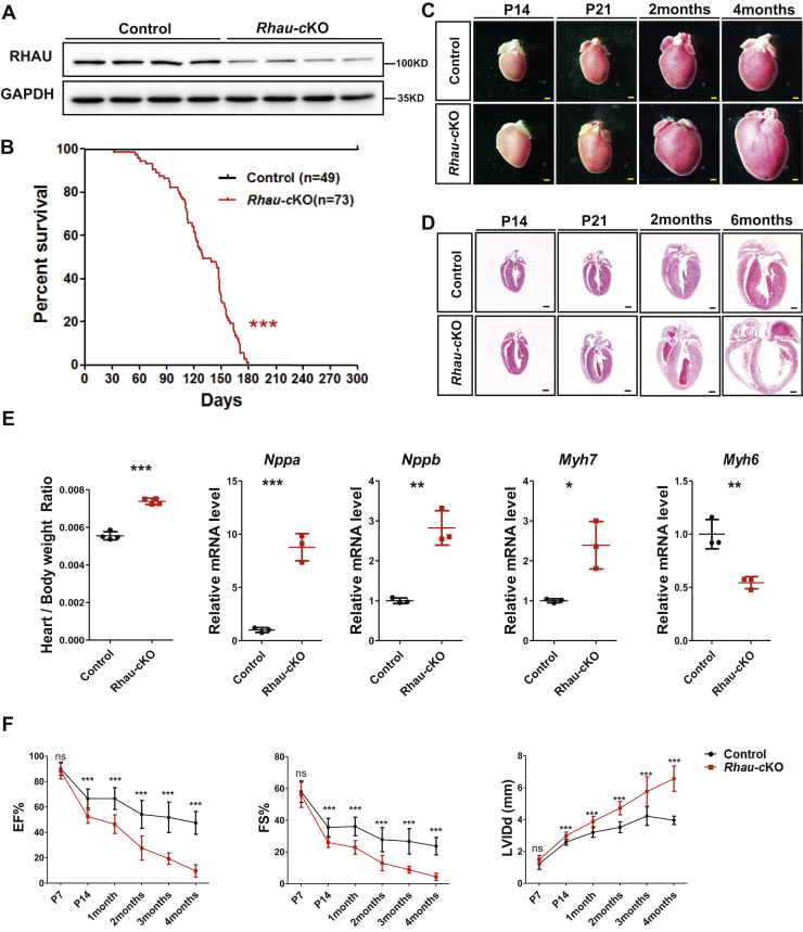
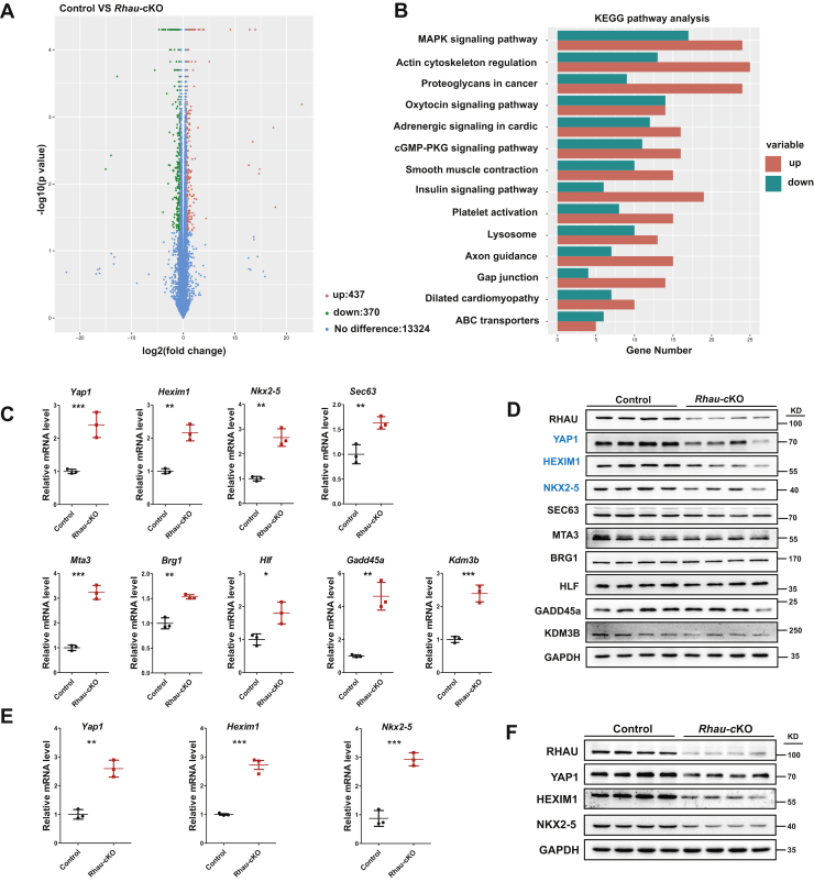
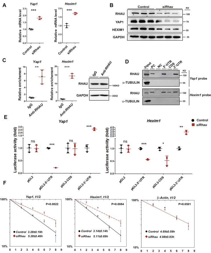
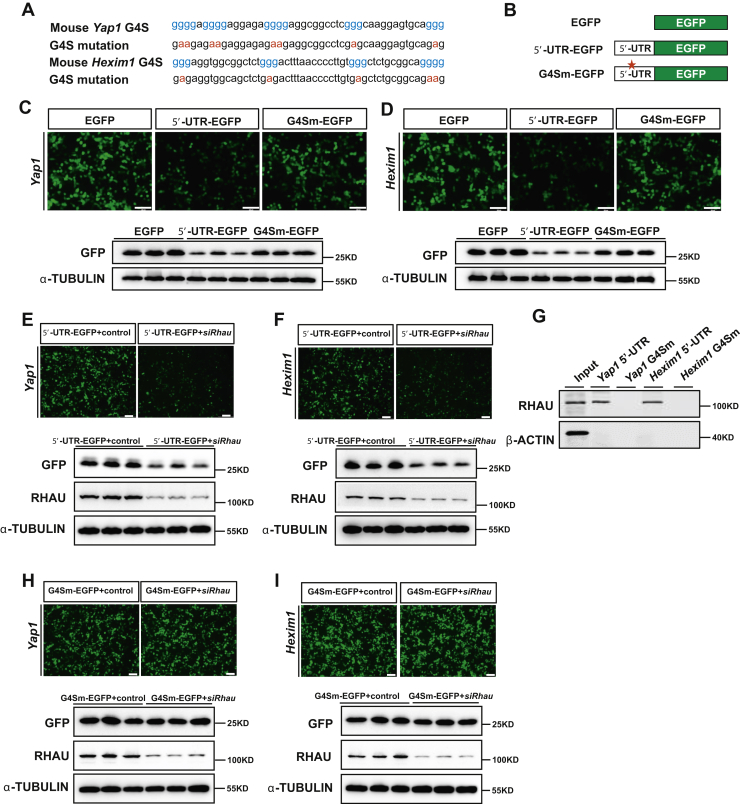
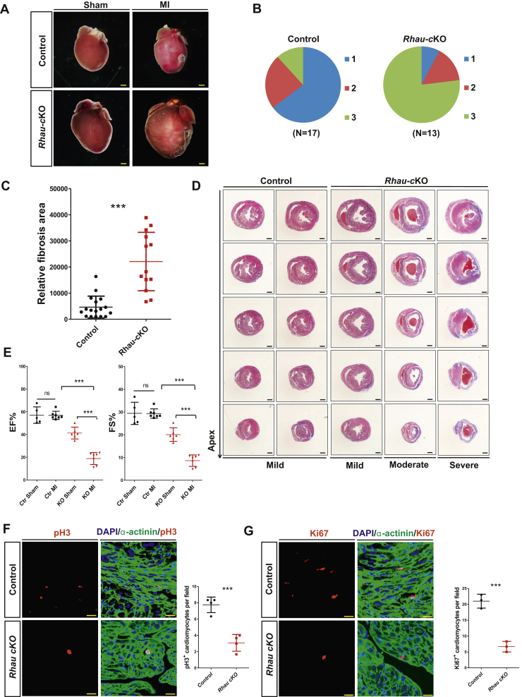
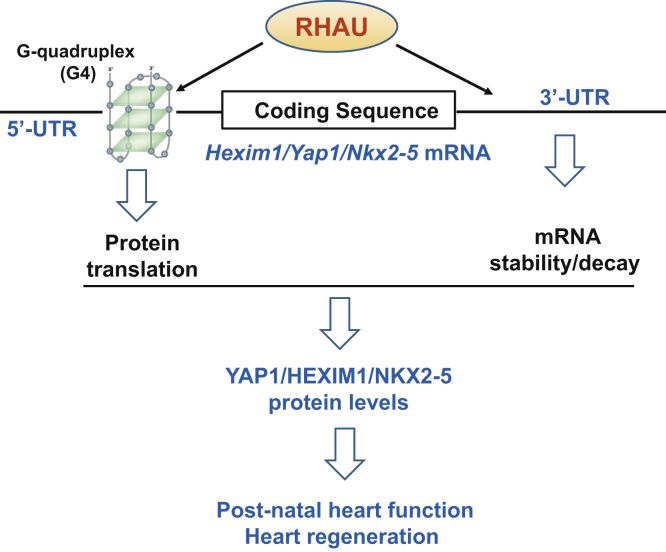
Post-transcriptional regulation of mRNA translation and stability is primarily achieved by RNA-binding proteins, which are of increasing importance for heart function. Furthermore, G-quadruplex (G4) and G4 resolvase activity are involved in a variety of biological processes. However, the role of G4 resolvase activity in heart function remains unknown. The present study aims to investigate the role of RNA helicase associated with adenylate- and uridylate-rich element (RHAU), an RNA-binding protein with G4 resolvase activity in postnatal heart function through deletion of Rhau in the cardiomyocytes of postnatal mice. RHAU-deficient mice displayed progressive pathological remodeling leading to heart failure and mortality and impaired neonatal heart regeneration. RHAU ablation reduced the protein levels but enhanced mRNA levels of Yap1 and Hexim1 that are important regulators for heart development and postnatal heart function. Furthermore, RHAU was found to associate with both the 5' and 3' UTRs of these genes to destabilize mRNA and enhance translation. Thus, we have demonstrated the important functions of RHAU in the dual regulation of mRNA translation and stability, which is vital for heart physiology.

Keywords: HEXIM1; RHAU; RNA-binding protein; YAP1; heart; mRNA; regeneration.


Conflict of interest statement

Conflict of interest The authors declare that they have no conflicts of interest with the contents of this article.

Figures

Figure 1
Figure 1
Rhau deletion induced dilated cardiomyopathy and heart failure.A, western blotting analysis confirmed effective Rhau deletion in Rhau-cKO (α-MHC-Cre;RhauF/F) mouse hearts at P7. Littermates (RhauF/F) were used as controls. GAPDH was used as a loading control. B, survival curves. N = 49 for control (RhauF/F) mice and n = 73 for Rhau-cKO (α-MHC-Cre;RhauF/F) mice. C, heart morphology of control (RhauF/F) mice and Rhau-cKO (α-MHC-Cre;RhauF/F) mice at P14, P21, 2 months, and 4 months. The scale bar represents 0.5 mm. D, histological analysis for control (RhauF/F) mice and Rhau-cKO (α-MHC-Cre;RhauF/F) mice at P14, P21, 2 months, and 6 months. The scale bar represents 0.5 mm. E, heart/body weight ratio analysis (n = 4) and quantitative RT-PCR (n = 3) analysis of cardiac hypertrophic markers in control (RhauF/F) mice and Rhau-cKO (α-MHC-Cre;RhauF/F) mice at P21. F, echocardiographic analysis of control (RhauF/F) mice and Rhau-cKO (α-MHC-Cre;RhauF/F) mice at various ages. EF, FS, and LVIDd were calculated according to the guidelines accompanying the Vevo 770 UBM system. Numbers of mice are as follows. Control group: P7 (n = 10), P14 (n = 14), 1 month (n = 9), 2 months (n = 11), 3 months (n = 9), and 4 months (n = 9); Rhau-cKO mice: P7 (n = 13), P14 (n = 11), 1 month (n = 9), 2 months (n = 11), 3 months (n = 9), and 4 months (n = 9). EF, ejection fraction; FS, fractional shortening; LVIDd, left ventricular internal diameter at end diastole; ns, not significant; RHAU, RNA helicase associated with adenylate- and uridylate-rich element.
Figure 2
Figure 2
Rhau deletion results in disrupted mRNA and protein levels of YAP1 and HEXIM1.A, volcano plot of RNA-Seq analysis. RNA was isolated from the hearts of control (RhauF/F) and Rhau-cKO (α-MHC-Cre;RhauF/F) mice at P10. Differentially expressed genes were filtered with fold change ≥1.5 and p < 0.05. B, KEGG pathway analysis of differentially expressed genes identified from RNA-Seq. C, quantitative RT-PCR analysis of candidate genes in the hearts of control (RhauF/F) mice and Rhau-cKO (α-MHC-Cre;RhauF/F) mice at P10. N = 3 for each group. D, western blotting analysis of candidate genes in the hearts of control mice and Rhau-cKO mice at P10. GAPDH was used as loading controls. E, quantitative RT-PCR analysis to detect Yap1, Hexim1, and Nkx2-5 mRNA levels. RNA was isolated from the hearts of control (RhauF/F) and Rhau-cKO (α-MHC-Cre;RhauF/F) mice at P21. N = 3 for each group. F, western blotting analysis examined the protein levels of YAP1, HEXIM1, and NKX2-5 in the hearts of control (RhauF/F) mice and Rhau-cKO (α-MHC-Cre;RhauF/F) mice at P21. GAPDH was used as loading controls. KEGG, Kyoto Encyclopedia of Genes and Genomes; MAPK, mitogen-activated protein kinase; PKG, protein kinase G.
Figure 3
Figure 3
RHAU regulates the stability of Yap1 and Hexim1 mRNAs.A, quantitative RT-PCR analysis to measure the mRNA levels of Yap1 and Hexim1 in control and Rhau knockdown H9C2 cells. Rhau was knocked down by transfection of siRNA targeting Rhau mRNAs, and control cells were transfected with a negative control siRNA. Seventy-two hours after transfection, cells were harvested. A half of the cells were used for RNA isolation, and the other half of the cells were used for Western blotting analysis. N = 3 for each group. B, qestern blotting analysis to detect the protein levels of RHAU, YAP1, and HEXIM1 in control and Rhau knockdown H9C2 cells. Protein was extracted from the cells described in (A). GAPDH was used as loading controls. C, ribonucleoprotein immunoprecipitation assays examined the endogenous association of RHAU with Yap1 and Hexim1 mRNA in H9C2 cells. The relative enriched levels of Yap1 and Hexim1 mRNA were determined by quantitative RT-PCR. The Gapdh transcripts were used as internal controls, and IgG antibody was used as negative controls. Western blotting analysis showed the enrichment of RHAU protein levels in the samples. N = 3 for each group. D, RNA pull-down assays were performed to assess the association of RHAU with biotinylated 5'-UTRs, CDSs, and 3'-UTRs of Yap1 and Hexim1 mRNA. The biotinylated 3'-UTR of Nkx2-5 mRNA was used as a positive control and biotinylated p27-CDS as a negative control. E, luciferase reporter assays explored the effects of RHAU binding to specific fragments of the Yap1 and Hexim1 mRNAs on Rhau knockdown in H9C2 cells. Rhau was knocked down by transfection of siRNA targeting Rhau mRNAs, and control cells were transfected with a negative control siRNA. For each column, n = 3. F, half-lives of the endogenous mRNAs of Yap1 and Hexim1 were examined in Rhau knockdown H9C2 cells and control (transfected with a negative control siRNA) cells. The β-actin mRNA was used as an internal control. For each time point, n = 3. IgG, immunoglobulin G; RHAU, RNA helicase associated with adenylate- and uridylate-rich element.
Figure 4
Figure 4
RHAU promoted the translation of Yap1 and Hexim1 mRNAs.A, potential G4-forming sequences (G4S) and their corresponding mutations in the 5'-UTRs of Yap1 and Hexim1 mRNA are depicted. B, schematic representation of the chimeric EGFP vector structure. EGFP represents pEGFP-N1 blank vector; 5'-UTR-EGFP represents chimeric pEGFP-N1 vector flanked with 5'-UTR of Yap1 or Hexim1 mRNA, and G4Sm-EGFP represents chimeric pEGFP-N1 vector flanked with G4S-mutated 5'-UTR of Yap1 or Hexim1 mRNA. C and D, representative images show the intensity of chimeric GFP reporters for Yap1 and Hexim1 followed by Western blotting analysis to confirm the GFP protein levels. α-Tubulin was used as a loading control. The scale bar represents 200 μm. E and F, representative images show that Rhau knockdown reduced the activity of chimeric GFP reporters for Yap1 and Hexim1 followed by Western blotting analysis. α-Tubulin was used as a loading control. The scale bar represents 200 μm. G, RNA pull-down assays were performed to assess the association of RHAU with biotinylated 5'-UTRs and G4S-mutated 5'-UTRs. H and I, representative images show the activity of G4Sm-EGFP reporters for Yap1 and Hexim1 in response to Rhau knockdown and Western blotting analysis. α-Tubulin was used as a loading control. The scale bar represents 200 μm. EGFP, enhanced GFP; RHAU, RNA helicase associated with adenylate- and uridylate-rich element.
Figure 5
Figure 5
Rhau deletion impairs heart regeneration in the neonatal mice.A, representative images from morphological analysis of control (RhauF/F) and Rhau-cKO (α-MHC-Cre;RhauF/F) mice after MI surgery or a sham operation. Four weeks after MI surgery or sham operation (performed at P5), the hearts were collected for analysis. The scale bar represents 0.5 mm. B, heart regeneration capability scoring of control (RhauF/F) mice (n = 17) and Rhau-cKO (α-MHC-Cre;RhauF/F) mice (n = 13). The regeneration capability was grouped into three categories: category 1 indicated complete regeneration, category 2 represented partial regeneration, and category 3 indicated a total blockade of regeneration. C, quantification of fibrotic scar areas for control (RhauF/F) mice (n = 17) and Rhau-cKO (α-MHC-Cre;RhauF/F) mice (n = 13) 4 weeks after MI surgery. D, representative images from Masson's trichrome staining of heart sections for control (RhauF/F) mice and Rhau-cKO (α-MHC-Cre;RhauF/F) mice 4 weeks after MI surgery. The scale bar represents 0.5 mm. E, Echocardiography tests to examine the cardiac function of control (RhauF/F) mice and Rhau-cKO (α-MHC-Cre;RhauF/F) mice after MI or sham operation. Four weeks after MI surgery or sham operation (performed at P5), the mice were used for analysis. EF and FS were calculated according to the guidelines. For sham group, n = 5 and n = 6 for control and Rhau-cKO mice, respectively; for MI group, n = 7 for control or Rhau-cKO mice. FG, immunofluorescence staining of pH3 (F) (n = 4) and Ki67 (G) (n = 3) labeled the proliferating cardiomyocytes after MI surgery and data quantification. One week after MI surgery performed at P5, the hearts of control (RhauF/F) mice and Rhau-cKO (α-MHC-Cre;RhauF/F) mice were collected for analysis. pH3 and Ki67 marked the proliferating cells, α-actinin marked the cardiomyocytes, and DAPI marked all the nuclei. The average number of proliferating cardiomyocytes from three fields per heart section was used for quantification. The scale bar represents 200 μm. DAPI, 4′,6-diamidino-2-phenylindole; EF, ejection fraction; FS, fractional shortening; MI, myocardial infarction; ns, not significant.
Figure 6
Figure 6
Working model of RHAU sustains postnatal heart function and neonatal regeneration through modulating target mRNA molecules. RHAU associates with both the 5'- and 3'-UTRs of the mRNA molecules of Yap1, Hexim1, and Nkx2-5 to regulate mRNA translation and stability. The proteins of YAP1, HEXIM1, and NKX2-5 are important regulators for heart function and regeneration. RHAU, RNA helicase associated with adenylate- and uridylate-rich element.

Similar articles

Cited by

References

    1. Krebs J.E., Lewin B., Goldstein E.S., Kilpatrick S.T. Jones & Bartlett Publishers, Burlington, MA; 2014. Lewin's Genes XI.
    1. Alberts B. Garland Science, New York, NY; 2017. Molecular Biology of the Cell.
    1. Lee T.I., Young R.A. Transcription of eukaryotic protein-coding genes. Annu. Rev. Genet. 2000;34:77–137. - PubMed
    1. Filipowicz W., Bhattacharyya S.N., Sonenberg N. Mechanisms of post-transcriptional regulation by microRNAs: are the answers in sight? Nat. Rev. Genet. 2008;9:102–114. - PubMed
    1. Jaenisch R., Bird A. Epigenetic regulation of gene expression: how the genome integrates intrinsic and environmental signals. Nat. Genet. 2003;33 Suppl:245–254. - PubMed

Publication types

MeSH terms