. 2021 Jan 8;12(1):174.
doi: 10.1038/s41467-020-20447-y.

Identification of a subset of immunosuppressive P2RX1-negative neutrophils in pancreatic cancer liver metastasis

Affiliations

Identification of a subset of immunosuppressive P2RX1-negative neutrophils in pancreatic cancer liver metastasis

Xu Wang et al. Nat Commun. .

Abstract

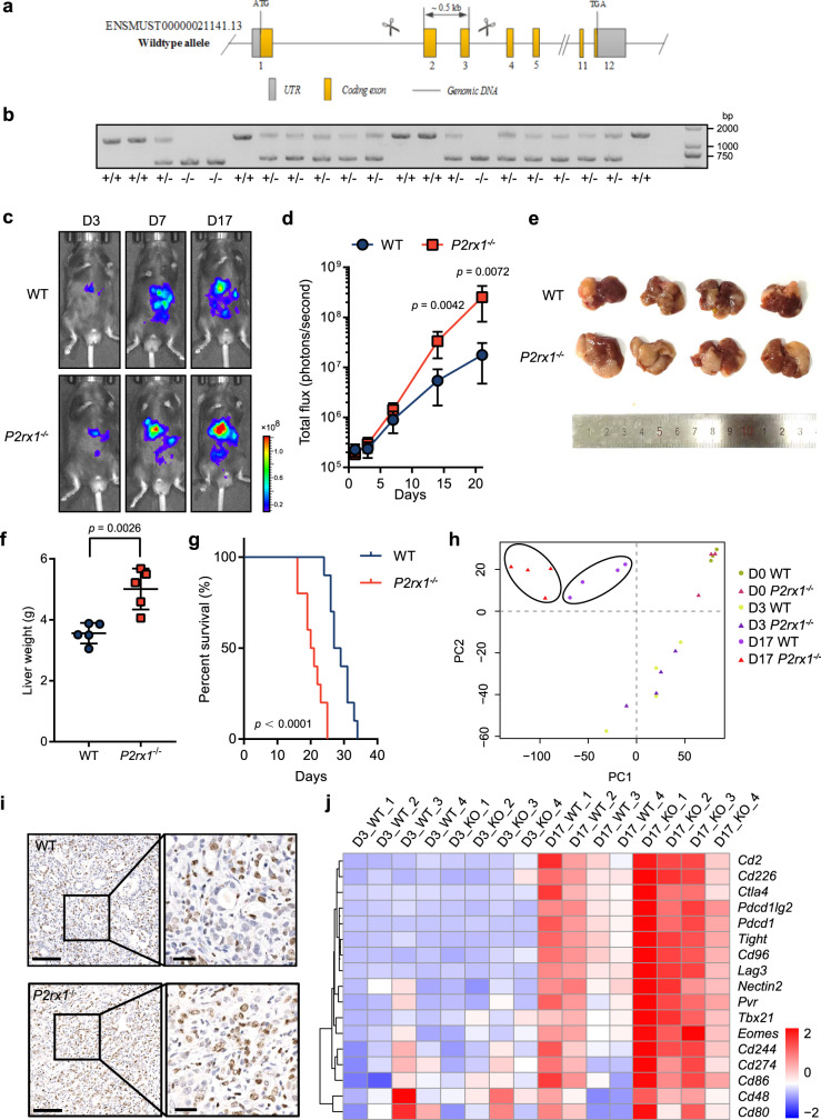
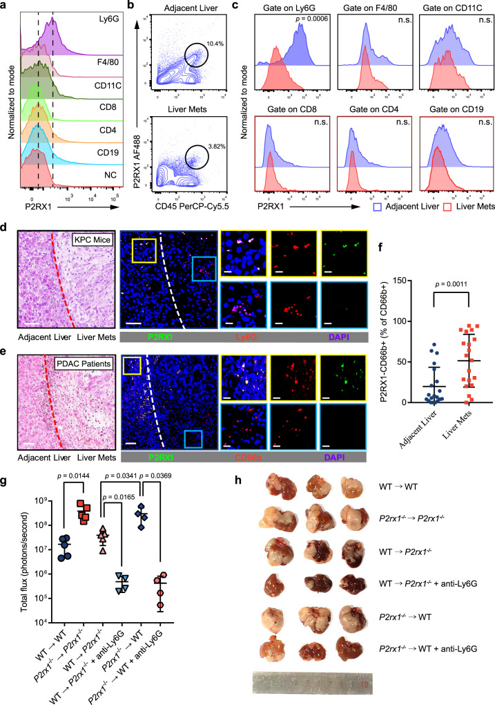
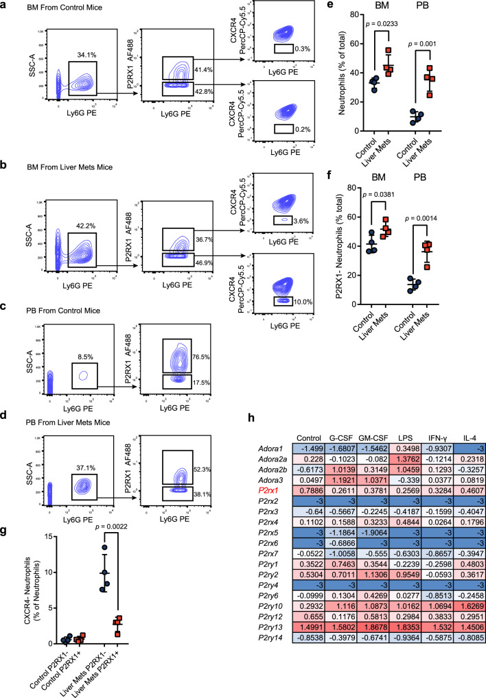
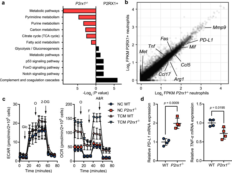
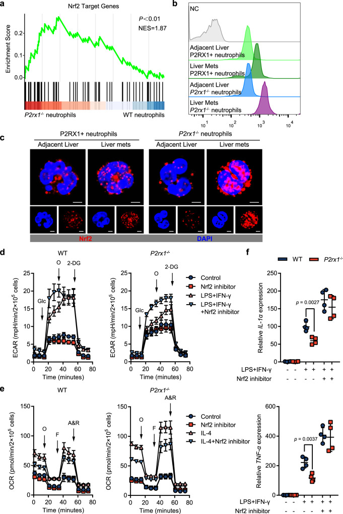
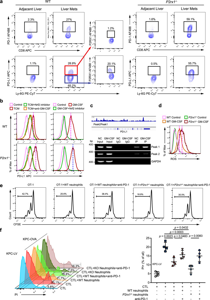
The immunosuppressive microenvironment that is shaped by hepatic metastatic pancreatic ductal adenocarcinoma (PDAC) is essential for tumor cell evasion of immune destruction. Neutrophils are important components of the metastatic tumor microenvironment and exhibit heterogeneity. However, the specific phenotypes, functions and regulatory mechanisms of neutrophils in PDAC liver metastases remain unknown. Here, we show that a subset of P2RX1-negative neutrophils accumulate in clinical and murine PDAC liver metastases. RNA sequencing of murine PDAC liver metastasis-infiltrated neutrophils show that P2RX1-deficient neutrophils express increased levels of immunosuppressive molecules, including PD-L1, and have enhanced mitochondrial metabolism. Mechanistically, the transcription factor Nrf2 is upregulated in P2RX1-deficient neutrophils and associated with PD-L1 expression and metabolic reprogramming. An anti-PD-1 neutralizing antibody is sufficient to compromise the immunosuppressive effects of P2RX1-deficient neutrophils on OVA-activated OT1 CD8+ T cells. Therefore, our study uncovers a mechanism by which metastatic PDAC tumors evade antitumor immunity by accumulating a subset of immunosuppressive P2RX1-negative neutrophils.


Conflict of interest statement

The authors declare no competing interests.

Figures

Fig. 1
Fig. 1. Reduced P2RX1 is associated with an immunosuppressive microenvironment in clinical PDAC liver metastases.
a Volcano plots of differential gene expression in 145 primary PDAC, 46 adjacent pancreases, 25 liver metastases and 27 adjacent livers. Red dots represent upregulated immune-related genes, and blue dots represent downregulated immune-related genes. b Immunome analyses of 26 infiltrating immune cell types in adjacent pancreas, primary PDAC, adjacent liver tissue and metastatic PDAC samples. c GO Biological Process analyses of differentially expressed genes between adjacent liver tissue and metastatic PDAC samples. d Expression analyses of P2RX1 in the adjacent pancreas, primary PDAC, adjacent liver tissue and metastatic PDAC samples from the GSE71729 and Renji cohorts. e Correlation analyses between P2RX1 and immune checkpoint molecules in metastatic PDAC samples. Bars represent mean ± standard deviation in (d). *P < 0.05, **P < 0.01, and ***P < 0.001, by one-way ANOVA and Tukey’s multiple comparisons test (d left), or Student’s t test (d right). Source data are provided as a Source data file.
Fig. 2
Fig. 2. P2RX1 deficiency promotes PDAC liver metastases and upregulates immune exhaustion-related gene expression.
a, b P2rx1−/− mice were generated using CRISPR/Cas9 system. Schematic diagram was shown in (a) and genotyping results were shown in (b) (representative result from three independent experiments). c, d KPC cells were intrasplenically injected to seed livers of WT and P2rx1−/− mice, and in vivo imaging was performed at sequential times. Representative images are shown in (c), and quantitative results are shown in (d) (n = 6 per group, three independent experiments). e Representative images of liver metastatic samples harvested at day 17. f Liver weight of liver metastatic samples was measured at day 17 (n = 5 per group, two independent experiments). g Survival analysis of liver metastatic WT or P2rx1−/− mice within a duration of 5 weeks (n = 10 per group, two independent experiments). h Normal liver (D0) and two sequential stages (D3 and D17) of liver metastases in WT and P2rx1−/− mice were harvested for RNA-seq, and PCA analyses were performed (n = 3 for D0, n = 4 for D3 and D17). i Representative Ki67 immunohistochemical staining of WT and P2rx1−/− liver metastases at day 17 (n = 4 per group, two independent experiments). 100 μm of scale bar for low power fields, 25 μm of scale bar for high power fields. j Heatmap of immune checkpoint molecules in liver metastases of WT or P2rx1−/− mice at D3 and D17 (n = 4 per group). Bars represent mean ± standard deviation in (d, f). P values are derived from two-sided Student’s t test (d, f), or log-rank test (g). Source data are provided as a Source data file.
Fig. 3
Fig. 3. P2RX1− neutrophils accumulate in the microenvironment of PDAC liver metastases and promote metastatic tumor growth.
a Single cell suspension was obtained from mouse spleen and expression of P2RX1 was determined in indicated immune cell types (n = 3 per group, two independent experiments). Left dotted line indicates the mean of negative control (NC) (secondary antibody only), and right dotted line indicates the mean of Ly6G+ cells. b KPC cells were intrasplenically injected to seed livers of WT mice. A single cell suspension was obtained from liver metastases and adjacent liver tissues of WT mice at day 17. The frequency of CD45+P2RX1+ cells was determined by flow cytometry (n = 4 per group, three independent experiments). c KPC cells were intrasplenically injected to seed livers of WT mice. Immune cells were enriched from single cell suspension of liver metastases and adjacent liver tissues at day 17. P2RX1 expression in the indicated immune cell types was determined by flow cytometry (n = 4 per group, three independent experiments). d Representative images of H&E staining and immunofluorescence staining of P2RX1 (Green), Ly6G (Red) and DAPI (Blue) in KPC mice spontaneous liver metastases (representative results from six independent experiments). 50 μm scale bar for low power fields and 20 μm scale bar for high power fields. e, f H&E staining and immunofluorescence staining of P2RX1 (green), CD66b (red) and DAPI (blue) in a total of 20 clinical PDAC liver metastasis samples were performed. Representative images are shown in (e), and the percentages of P2RX1-CD66b+ cells are shown in (f). 50 μm scale bar for low power fields and 20 μm scale bar for high power fields. g, h KPC cells were intrasplenically injected to seed livers of BM chimeras: WT → WT, P2rx1−/− → P2rx1−/−, WT → P2rx1−/−, and P2rx1−/− → WT. Neutrophils were depleted in WT → P2rx1−/− and P2rx1−/− → WT mice by intraperitoneal injection of anti-Ly6G (clone 1A8) antibody. At day 17, liver metastases were analyzed by in vivo imaging (g, n = 5 for WT → WT, P2rx1−/−P2rx1−/− and WT → P2rx1−/− groups, n = 4 for WT → P2rx1−/− + anti-Ly6G, P2rx1−/− → WT and P2rx1−/− → WT + anti-Ly6G groups, two independent experiments), and representative images of liver metastatic samples were shown (h). Bars represent mean ± standard deviation in (f, g). P values are derived from two-sided Student’s t test (c, f), or one-way ANOVA and Tukey’s multiple comparisons test (g). Source data are provided as a Source data file.
Fig. 4
Fig. 4. PDAC liver metastasis systematically mobilizes and recruits P2RX1− neutrophils.
ag KPC cell was intrasplenically injected to seed livers of WT mice. Bone marrow (BM) and peripheral blood (PB) were obtained at day 17. Frequencies of neutrophils, P2RX1− neutrophils, and CXCR4+ neutrophils were determined by flow cytometry and quantitative results were shown (n = 4 per group, three independent experiments). h Bone marrow neutrophils were isolated from WT mice and stimulated with indicated stimulus. RNA-seq were performed and Log2 (FPKM+0.001) value of purinergic receptors were shown (n = 1 per group). Bars represent mean ± standard deviation in (eg). P values are derived from two-sided Student’s t test (eg). Source data are provided as a Source data file.
Fig. 5
Fig. 5. Characterizing the immune and metabolic features of P2RX1− neutrophils in the microenvironment of PDAC liver metastases.
a, b KPC cells were intrasplenically injected to seed livers of WT and P2rx1−/− mice. A single cell suspension was obtained from liver metastases at day 17. Then, P2RX1+ neutrophils were purified from WT mice, and P2rx1−/− neutrophils were purified from P2rx1−/− mice for RNA sequencing. The results of KEGG analysis are shown in (a), and comparative expression of genes is shown in (b) (n = 1 per group). c Bone marrow neutrophils were isolated from WT and P2rx1−/− mice and stimulated with tumor conditioned medium (TCM). The ECAR and OCR were then measured by a Seahorse assay in (c), and PD-L1 and TNF-α were detected by RT-qPCR in (d) (n = 4 per group, two independent experiments). Glc, glucose; O (ECAR), oligomycin; 2-DG, 2-deoxyglucose; O (OCR), oligomycin; F, FCCP (carbonyl cyanide 4-[trifluoromethoxy] phenylhydrazone); A & R, antimycin A and rotenone. Bars represent mean ± standard deviation in (c, d). P values are derived from two-sided Student’s t test (d). Source data are provided as a Source data file.
Fig. 6
Fig. 6. Upregulated Nrf2 is essential for shaping the immune-tolerant phenotype of P2RX1− neutrophils.
a Gene set enrichment analysis comparing RNA-seq data of P2RX1+ neutrophils and P2rx1−/− neutrophils based on Nrf2 target genes. The p value and normalized enrichment score (NES) were shown. b, c Single-cell suspensions were obtained from liver metastases of WT and P2rx1−/− mice at day 17. Intracellular Nrf2 was stained and detected by flow cytometry (b) or laser scanning confocal microscopy (c) in WT P2RX1+ neutrophils and P2rx1−/− neutrophils (n = 4 per group, three independent experiments). The scale bar is 2.5 μm. d Bone marrow neutrophils were isolated from WT and P2rx1−/− mice and stimulated with LPS + IFN-γ in the presence or absence of a Nrf2 inhibitor. The ECAR was then measured by a Seahorse assay (n = 4 per group, two independent experiments). Glc, glucose; O, oligomycin; 2-DG, 2-deoxyglucose. e Bone marrow neutrophils were isolated from WT and P2rx1−/− mice and stimulated with IL-4 in the presence or absence of a Nrf2 inhibitor. The OCR was then measured by a Seahorse assay (n = 4 per group, two independent experiments). O, oligomycin; F, FCCP (carbonyl cyanide 4-[trifluoromethoxy] phenylhydrazone); A & R, antimycin A and rotenone. f Bone marrow neutrophils were isolated from WT or P2rx1−/− mice and stimulated with LPS + IFN-γ and an inhibitor Nrf2 inhibitor. IL-1β and TNF-α were determined by RT-qPCR (n = 4 per group, three independent experiments). Bars represent mean ± standard deviation in (df). P values are derived from permutation test (a), two-sided Student’s t test (f). Source data are provided as a Source data file.
Fig. 7
Fig. 7. P2RX1− neutrophils facilitate an immunosuppressive microenvironment in PDAC liver metastases by upregulating PD-L1 expression.
a A single cell suspension was obtained from liver metastases of WT and P2rx1−/− mice at day 17. Flow cytometry was performed to detect the frequency of CD8+PD-1+ (upper) and Ly6G+PD-L1 + or Ly6G+PD-L1− (lower) cells. P2RX1 expression was further determined in Ly6G+PD-L1+ and Ly6G+PD-L1− cells from WT mice (n = 4 per group, three independent experiments). b Bone marrow neutrophils were isolated from WT and P2rx1−/− mice and stimulated with tumor conditioned medium (TCM) or GM-CSF in the presence of a Nrf2 inhibitor or anti-GM-CSF neutralizing antibody. PD-L1 expression was detected by flow cytometry (n = 4 per group, three independent experiments). c Integrative Genomics Viewer (IGV) was used to predict the two peaks that PD-L1 gene might be mediated by Nrf2 (upper). Bone marrow neutrophils were isolated from WT and P2rx1−/− mice and stimulated with GM-CSF. Binding of PD-L1 gene by Nrf2 was detected by ChIP-PCR (n = 3 per group, two independent experiments). d Bone marrow neutrophils were isolated from WT and P2rx1−/− mice and stimulated with GM-CSF. Intracellular ROS was detected by flow cytometry (n = 4 per group, three independent experiments). e Antigen activated CTLs was co-cultured with GM-CSF primed WT or P2rx1−/− neutrophils. Cell proliferation was analyzed with CSFE staining in the presence or absence of anti-PD-1 neutralizing antibody (n = 4 per group, three independent experiments). f KPC cells were transfected with empty lentiviral vector (KPC-LV) or OVA (KPC-OVA). Antigen-activated CTLs were co-cultured with KPC-LV or KPC-OVA cells in the presence of GM-CSF-primed WT or P2rx1−/− neutrophils. Cytotoxicity was determined in the presence or absence of anti-PD-1 neutralizing antibody by counting the number of PI+ cells (n = 4 per group, three independent experiments). Bars represent mean ± standard deviation in (f). P values are derived from one-way ANOVA and Tukey’s multiple comparisons test (f). Source data are provided as a Source data file.

Similar articles

Cited by

References

    1. Wolfgang CL, et al. Recent progress in pancreatic cancer. CA Cancer J. Clin. 2013;63:318–348. doi: 10.3322/caac.21190. - DOI - PMC - PubMed
    1. Roe JS, et al. Enhancer Reprogramming Promotes Pancreatic Cancer Metastasis. Cell. 2017;170:875–888 e820. doi: 10.1016/j.cell.2017.07.007. - DOI - PMC - PubMed
    1. Makohon-Moore AP, et al. Limited heterogeneity of known driver gene mutations among the metastases of individual patients with pancreatic cancer. Nat. Genet. 2017;49:358–366. doi: 10.1038/ng.3764. - DOI - PMC - PubMed
    1. Moffitt RA, et al. Virtual microdissection identifies distinct tumor- and stroma-specific subtypes of pancreatic ductal adenocarcinoma. Nat. Genet. 2015;47:1168–1178. doi: 10.1038/ng.3398. - DOI - PMC - PubMed
    1. Goehrig D, et al. Stromal protein betaig-h3 reprogrammes tumour microenvironment in pancreatic cancer. Gut. 2019;68:693–707. doi: 10.1136/gutjnl-2018-317570. - DOI - PMC - PubMed

Publication types

MeSH terms