. 2021 Aug 26;20(1):108.
doi: 10.1186/s12943-021-01403-w.

DDX56 modulates post-transcriptional Wnt signaling through miRNAs and is associated with early recurrence in squamous cell lung carcinoma

Affiliations

DDX56 modulates post-transcriptional Wnt signaling through miRNAs and is associated with early recurrence in squamous cell lung carcinoma

Qingqing Wu et al. Mol Cancer. .

Abstract

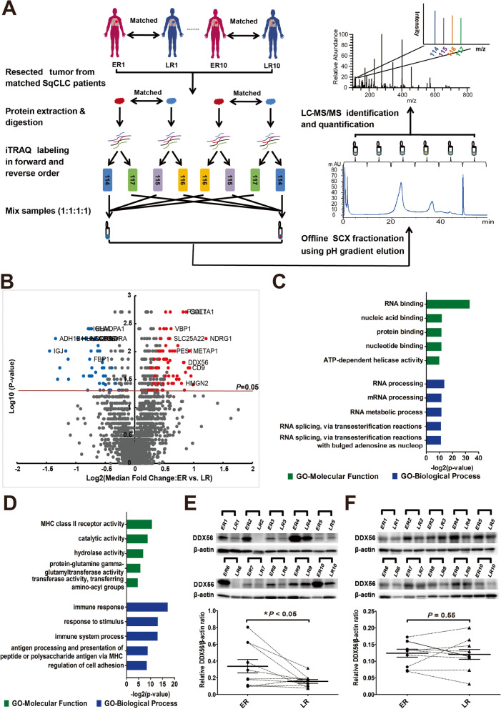
Background: Early recurrence is a major obstacle to prolonged postoperative survival in squamous cell lung carcinoma (SqCLC). The molecular mechanisms underlying early SqCLC recurrence remain unclear, and effective prognostic biomarkers for predicting early recurrence are needed.

Methods: We analyzed primary tumor samples of 20 SqCLC patients using quantitative proteomics to identify differentially-expressed proteins in patients who experienced early versus late disease recurrence. The expression and prognostic significance of DDX56 was evaluated using a SqCLC tumor tissue microarray and further verified using different online databases. We performed in vitro and in vivo experiments to obtain detailed molecular insight into the functional role of DDX56 in SqCLC.

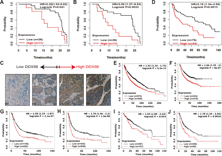
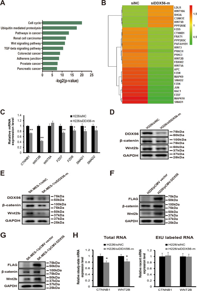
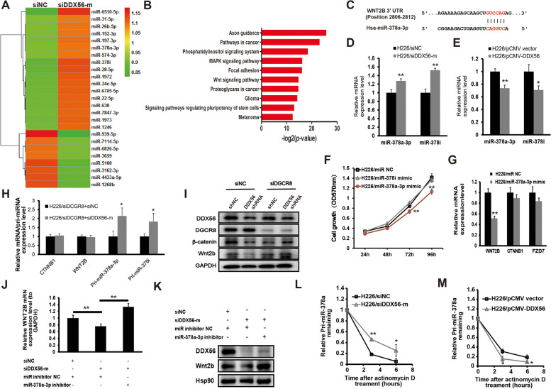
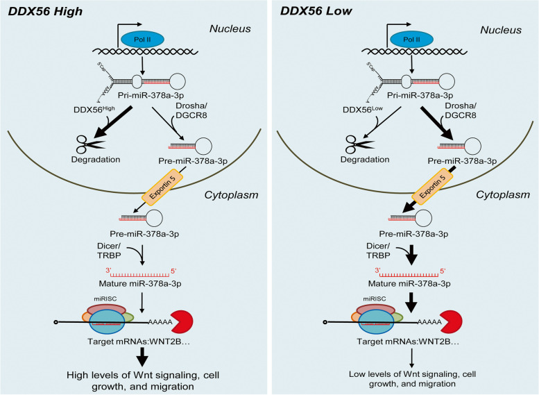
Results: We found that DDX56 exhibited increased expression in tumors of patients who experienced early versus late disease recurrence. Increased DDX56 expression in SqCLC tumors was subsequently confirmed as an independent prognostic factor of poor recurrence-free survival in independent SqCLC cohorts. Functionally, DDX56 promotes SqCLC cell growth and migration in vitro, and xenograft tumor progression in vivo. Mechanistically, DDX56 post-transcriptionally promotes expression of multiple Wnt signaling pathway-related genes, including CTNNB1, WNT2B, and represses a subset of miRNAs, including miR-378a-3p, a known suppressor of Wnt signaling. Detailed analysis revealed that DDX56 facilitated degradation of primary miR-378a, leading to down-regulation of mature miR-378a-3p and thus derepression of the target gene WNT2B.

Conclusion: We identified DDX56 as a novel independent prognostic biomarker that exerts its oncogenic effects through miRNA-mediated post-transcriptional regulation of Wnt signaling genes to promote early SqCLC recurrence. DDX56 may assist in identifying SqCLC patients at increased risk of early recurrence and who could benefit from Wnt signaling-targeted therapies.

Keywords: DDX56; Squamous cell lung cancer; Wnt signaling pathway; miRNA.


Conflict of interest statement

The authors declare that they have no competing interests.

The authors confirm that there is no conflict of interest related to this manuscript.

Figures

Fig. 1
Fig. 1
Identification of differentially-expressed proteins in primary squamous cell lung cancer (SqCLC) tissues of patients exhibiting early recurrence (ER) versus late recurrence (LR). a Experimental scheme of quantitative proteomic analysis. SqCLC tumor sample of patient with early recurrence (within 10 months) following surgery was paired with a SqCLC tumor sample of a patient with late recurrence (> 30 months) based on similar clinical and pathological features. After tissue lysis and tryptic digestion, each pair of peptide samples were labeled with iTRAQ reagents twice in two different orders (forward and reverse), and were mixed in a ratio of 1:1:1:1. Each mixed iTRAQ-labeled proteomic sample was fractionated into 6 fractions by SCX chromatography using a pH gradient elution, and then analyzed by LC–MS/MS. b Volcano plot constructed using median fold-change values and p-values to compare protein expression changes between ER and matched LR patients. Red dots represent significantly upregulated proteins and blue dots represent downregulated proteins (> 1.5-fold change in at least 4 sample sets and P < 0.05). The horizontal line represents a P value cut off < 0.05. P values were calculated using two-sided one sample Wilcoxon signed rank test. c and d Gene ontology (GO) classification of the 71 upregulated genes (c) and 60 downregulated genes (d) identified by proteomic analysis. Top 5 enriched GO terms of each category are shown. Green bars represent molecular function terms; blue bars represent biological process terms. The horizontal axis represents the –log2 (p-value). P < 0.05 was used as the threshold for selecting significant GO categories. e and f Western blots were performed to confirm altered DDX56 protein levels in tumor tissues as identified by mass spectrometry analysis, and to further analyze DDX56 protein levels in corresponding adjacent normal lung tissues from the same ER and matched LR patients. e Western blot analysis of DDX56 in tumor tissues from paired ER or LR SqCLC patients in cohort 1. Band intensity of DDX56 for the paired ER or LR tumor tissue samples is shown as quantified by densitometry and normalized to β-actin. f Western blot analyses of DDX56 in adjacent normal tissues from patients in cohort 1. Band intensity of DDX56 for the paired ER or LR patient adjacent normal tissue samples as shown in e quantified by densitometry and normalized to β-actin. Horizontal bars represent the median value with standard error of the mean. P value is determined by two-sided paired Wilcoxon signed-rank test. * P < 0.05
Fig. 2
Fig. 2
High DDX56 tumor expression is significantly associated with poor patient outcome in SqCLC and other cancers. a, b and c Immunohistochemical analysis showing that high DDX56 tumor protein level is associated with poor prognosis of SqCLC patients. Tissue microarrays (TMA) containing cores of 37 SqCLC were graded as exhibiting no, weak, moderate and strong DDX56 staining by a highly experienced pathologist blinded to all clinicopathologic data. Kaplan–Meier analysis comparing tumors graded as no or weak staining (low) versus those graded moderate and strong staining (high) showed that the overall survival (OS) (a) and recurrence-free survival (RFS) (b) of SqCLC patients with high DDX56 protein tumor expression is significantly lower than those with low DDX56 protein tumor expression. c Representative images of the different immunohistochemistry staining intensities. d Kaplan–Meier survival analysis of DDX56 in a published microarray gene expression dataset of 130 primary SqCLC tumor samples showed that high mRNA expression of DDX56 was significantly associated with poor OS of SqCLC patients. e-j Online survival analysis of the prognostic value of DDX56 mRNA expression in lung, gastric and liver cancers using KMplot (http://www.kmplot.com). DDX56 mRNA expression levels were shown to be significantly associated with OS (e) and progression-free survival (PFS) (f) in lung cancer patients (microarray data set, n = 1925). DDX56 mRNA expression levels were shown to be significantly associated with poor OS (g) and PFS (h) in gastric cancer patients (microarray data set, n = 875). DDX56 mRNA expression levels were shown to be significantly associated with OS (i) and PFS (j) in liver cancer patients (RNA-seq data set, n = 364)
Fig. 3
Fig. 3
Reduction of DDX56 inhibits SqCLC cell growth, migration and tumor progression in a xenograft mouse model of SqCLC. a and b Analysis of growth of H226 (a) and SK-MES-1 (b) cells transfected with DDX56-specific or control shRNAs using a crystal violet assay. Cell growth of H226 and SK-MES-1 was significantly inhibited in DDX56 shRNA-transfected cells compared to control shRNA (shNC)-transfected cells (n = 4). c and d Analysis of growth of H226 and SK-MES-1 cells transfected with pCMV-DDX56 or control pCMV vectors using a crystal violet assay. Cell growth of H226 and SK-MES-1 was significantly enhanced in pCMV-DDX56-transfected cells compared to pCMV vector-transfected cells (n = 4). e and f Analysis of migration ability of H226 cells transfected with DDX56 or control siRNAs using wound healing assay. (e) Wound healing assay of H226 cells transfected with DDX56-specific or control siRNAs. (f) Quantification of wound closure by measuring the wound width of the % of the closure of original wound in triplicate plates. g and h Analysis of migration ability of H226 (g) and SK-MES-1 (h) cells transfected with DDX56-specific or control siRNAs using a transwell assay. Representative images of crystal violet-stained migrated H226 (200X) and SK-MES-1 (400X) cells on the membrane (left panel). Quantification of cell migration expressed by cell counting. Columns represent mean (n = 4). i and j, NOG CIEA were injected subcutaneously with H226 cells transfected with shDDX56 (n = 10) or shNC (n = 10) and monitored for H226 tumor growth at day 10, 19, 29 and 33 after injection (i). The tumor-bearing mice were sacrificed on day 33 and their tumors dissected. Representative tumors from DDX56 shRNA (n = 5) and control shRNA (n = 4) xenografts (j). Data are presented as mean ± SD. Two-sided Student’s t-test was used to compare the data. (*p < 0.05; **p < 0.01, ***p < 0.001, ****p < 0.0001). All data are representative of three independent experiments
Fig. 4
Fig. 4
DDX56 post-transcriptionally regulates expression of the Wnt signaling pathway-related genes. a GO (KEGG) enrichment analysis of down-regulated mRNAs (> twofold) in a mRNA microarray experiment revealed that the KEGG terms “pathways in cancer” and “Wnt signaling pathway” were significantly enriched in down-regulated mRNAs in DDX56 siRNA- vs. control siRNA-transfected cells. b Heatmap showing WNT signaling-related genes differentially expressed (> twofold) between scramble siRNA (left) and DDX56 siRNA-treated (right) H226 cells in a mRNA microarray experiment. c qRT-PCR analysis of CTNNB1, WNT2B, WNT5A, FZD7, FZD6, SMAD1, and SMAD2 mRNA expression levels in DDX56-specific siRNA (siDDX56-mix) or negative control siRNA- (siNC) transfected H226 cells showed that DDX56 reduction significantly reduced mRNA expression of Wnt pathway-related genes CTNNB1, WNT2B, FZD7, FZD6, SMAD1, and SMAD2 except for WNT5A in H226 cells. d and e Western blot analysis of β-catenin, Wnt2b, and DDX56 protein expression level in H226 and SK-MES-1 cells transfected with DDX56-specific siRNAs or control siRNA, confirming reduced protein expression of β-catenin and Wnt2b following DDX56 reduction in H226 (d) and SK-MES-1 (e) cells. f and g Western blot analysis of the expression levels of β-catenin, Wnt2b and FLAG tag-fused DDX56 protein in H226 and SK-MES-1 cells transfected with pCMV-DDX56 or control pCMV vectors, confirming enhanced protein expression of β-catenin and Wnt2b following DDX56 overexpression in H226 (f) and SK-MES-1 (g) cells. GAPDH expression was used as a loading control. h Nascent transcript analysis showed no significant variation in the newly synthesized EtU-labeled RNA of CTNNB1 and WNT2B, whereas a significant decrease in the total (steady-state) RNA levels of CTNNB1 and WNT2B was observed after siRNA-mediated DDX56 knockdown in H226 cells. Gene expression levels were measured by qRT-PCR and standardized with GAPDH. Data are graphed as mean ± SD (n = 4). * P < 0.05
Fig. 5
Fig. 5
miR-378a-3p as a downstream effector of DDX56 involved in regulation of WNT2B expression in SqCLC cells. a Heatmap showing differentially-expressed miRNAs (≥ 1.2-fold) between scramble siRNA (left) and DDX56 siRNA-treated (right) H226 cells in a miRNA microarray experiment. b GO (KEGG) enrichment analysis of predicted targets of upregulated miRNAs (≥ 1.2-fold) in a miRNA microarray experiment revealed that the KEGG terms “pathways in cancer” and “Wnt signaling pathway” were also significantly enriched in predicted targets of up-regulated miRNAs in DDX56 siRNA- vs. control siRNA-transfected cells. c Predicted binding site of human miR-378a-3p to the 3’UTR of WNT2B by TargetScan. d and e qRT-PCR analysis of mature miRNA levels of miR-378a-3p and miR-378i in DDX56 silenced or overexpressed H226 cells compared to control cells. d H226 cells were transfected with DDX56-specific siRNA or control siRNA. e H226 cells were transfected with pCMV-DDX56 vector or pCMV empty vector. U6B was used for normalization of the mature miRNA expression data. (n = 3). f Analysis of growth of H226 cells transfected with miR-378a-3p mimic, miR-378i mimic or negative control miRNA mimic as determined by a crystal violet assay. (n = 4). g qRT-PCR analysis of WNT2B mRNA expression level in H226 cells transfected with miR-378a-3p mimic or control miRNA. GAPDH was used for normalization. (n = 3). h qRT-PCR analysis of mRNA expression of WNT2B and CTNNB1 and primary miRNA expression of miR-378a-3p and miR-378i in H226 cells co-transfected with DGCR8 siRNA and control siRNA or DGCR8 siRNA and DDX56 siRNA. GAPDH was used for normalization. i Representative Western blot analysis of Wnt2b and β-catenin protein expression levels in H226 cells co-transfected with control siRNA and control shRNA or control siRNA and DDX56 shRNA or DGCR8 siRNA and control shRNA or DGCR8 siRNA and DDX56 shRNA. GAPDH was used as a loading control. (n = 3). j qRT-PCR analysis of WNT2B mRNA expression level in H226 cells co-transfected with control miRNA and control siRNA or control miRNA and DDX56 siRNA or miR-378a-3p mimic and DDX56 siRNA. GAPDH was used for normalization. k Representative Western blot analysis of Wnt2b protein expression level in H226 cells co-transfected with control miRNA and control siRNA or control miRNA and DDX56 siRNA or miR-378a-3p mimic and DDX56 siRNA. Expression of a housekeeping gene, Hsp90 was used as a loading control. (n = 3). l and m Pri-miRNA stability assay carried out using H226 cells. l H226 cells were transfected with DDX56-specific siRNA (si-DDX56-mix) or control siRNA for 48 h, then treated with the transcription inhibitor actinomycin D. Primary miR378a were quantitated by qRT-PCR normalized to 18 s rRNA at 0, 3, and 6 h. (n = 3). m H226 cells were transfected with pCMV-DDX56 or empty pCMV vector for 48 h, then treated with the transcription inhibitor actinomycin D. Primary miR378a were quantitated by qRT-PCR, normalized to 18 s rRNA at 0, 3, and 6 h. (n = 3). Primary miR-378a expression level at 0 h was defined as 1. Each experiment was performed independently at least twice and results are presented as mean ± SD. Two-sided Student’s t-test was used to analyze the data. (*p < 0.05; **p < 0.01)
Fig. 6
Fig. 6
A proposed model for DDX56 regulation of miR-378a-3p and subsequent modulation of WNT signaling in SqCLC cells. A proposed working model showing the DDX56/miR-378a-3p/Wnt2b signaling axis and its roles in SqCLC cells with high (left) or low (right) DDX56 expression. Thick arrows indicate more flow; thin arrows indicate less flow

Similar articles

Cited by

References

    1. Torre LA, Bray F, Siegel RL, Ferlay J, Lortet-Tieulent J, Jemal A. Global cancer statistics, 2012. CA Cancer J Clin. 2015;65:87–108. doi: 10.3322/caac.21262. - DOI - PubMed
    1. Herbst RS, Heymach JV, Lippman SM. Lung cancer. N Engl J Med. 2008;359:1367–1380. doi: 10.1056/NEJMra0802714. - DOI - PMC - PubMed
    1. Heist RS, Sequist LV, Engelman JA. Genetic changes in squamous cell lung cancer: a review. J Thorac Oncol. 2012;7:924–933. doi: 10.1097/JTO.0b013e31824cc334. - DOI - PMC - PubMed
    1. Carnio S, Novello S, Papotti M, Loiacono M, Scagliotti GV. Prognostic and predictive biomarkers in early stage non-small cell lung cancer: tumor based approaches including gene signatures. Transl Lung Cancer Res. 2013;2:372–381. - PMC - PubMed
    1. Uramoto H, Tanaka F. Recurrence after surgery in patients with NSCLC. Transl Lung Cancer Res. 2014;3:242–249. - PMC - PubMed

Publication types

MeSH terms