. 2021 Aug 11;12(1):4852.
doi: 10.1038/s41467-021-25032-5.

USP12 downregulation orchestrates a protumourigenic microenvironment and enhances lung tumour resistance to PD-1 blockade

Affiliations

USP12 downregulation orchestrates a protumourigenic microenvironment and enhances lung tumour resistance to PD-1 blockade

Zhaojuan Yang et al. Nat Commun. .

Abstract

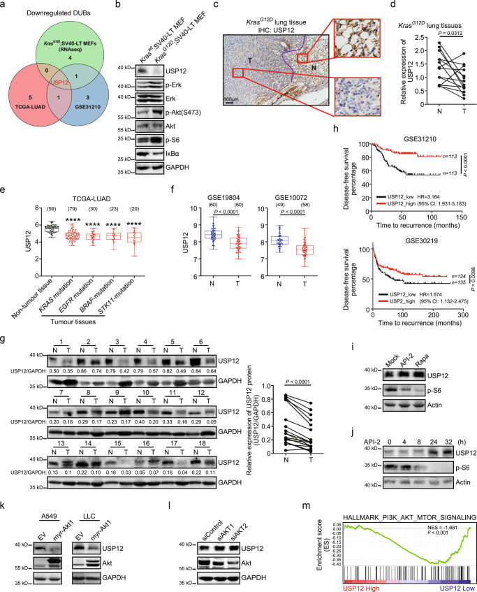
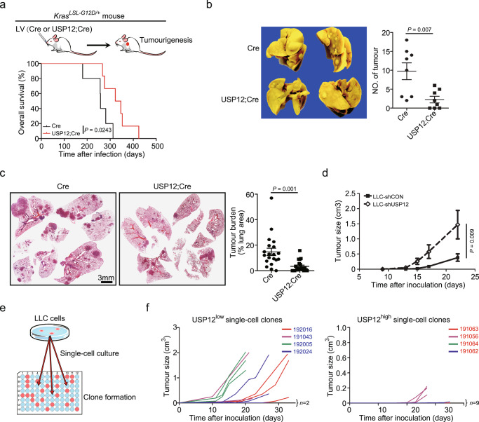
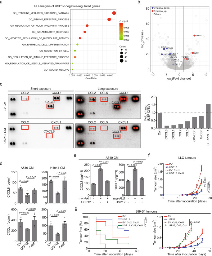
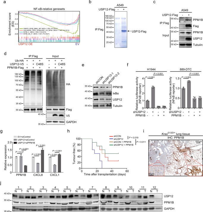
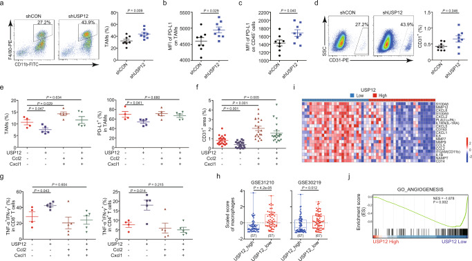
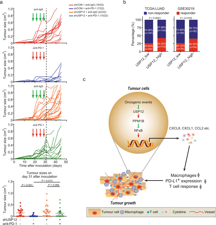
Oncogenic activation of KRAS and its surrogates is essential for tumour cell proliferation and survival, as well as for the development of protumourigenic microenvironments. Here, we show that the deubiquitinase USP12 is commonly downregulated in the KrasG12D-driven mouse lung tumour and human non-small cell lung cancer owing to the activation of AKT-mTOR signalling. Downregulation of USP12 promotes lung tumour growth and fosters an immunosuppressive microenvironment with increased macrophage recruitment, hypervascularization, and reduced T cell activation. Mechanistically, USP12 downregulation creates a tumour-promoting secretome resulting from insufficient PPM1B deubiquitination that causes NF-κB hyperactivation in tumour cells. Furthermore, USP12 inhibition desensitizes mouse lung tumour cells to anti-PD-1 immunotherapy. Thus, our findings propose a critical component downstream of the oncogenic signalling pathways in the modulation of tumour-immune cell interactions and tumour response to immune checkpoint blockade therapy.


Conflict of interest statement

The authors declare no competing interests.

Figures

Fig. 1
Fig. 1. USP12 expression is commonly downregulated in human NSCLC and KrasG12D-driven mouse lung tumours.
a Venn diagram showing the overlapped DUB genes that were significantly downregulated in cells with Kras activation and clinical NSCLCs. Data from our RNA-seq analysis of tumourigenic KrasG12D;SV40-LT MEFs vs. non-tumourigenic Kraswt;SV40-LT MEFs, and from TCGA and GSE31210. b Western blotting showing indicated protein levels in MEFs with or without KrasG12D expression. c, d Representative immunohistochemistry (IHC) staining (c, representative images of three biologically independent mice) and USP12 mRNA levels (d) in lung tissues from KrasLSL-G12D/+ mice 2 months after Ad-Cre infection. 2-tailed paired t-test. T: tumour; N: non-tumour tissue. e Boxplot showing USP12 levels in NSCLC patients with the mutations of KRAS, EGFR, BRAF and STK11 genes. The data were obtained from the TCGA-LUAD database. ****P < 0.0001 vs. non-tumour tissue by 2-tailed unpaired t-test. f Boxplots of USP12 gene expression in the NSCLC samples from the GSE19804 and GSE10072 databases. 2-tailed unpaired t-test. g USP12 protein levels in human NSCLC samples. The quantification analysis is shown on the right (n = 18). 2-tailed paired t-test. h Kaplan-Meier plots showing the disease-free survival of NSCLC patients based on USP12 expression. Two-sided log-rank test. HR: hazard ratios; CI: confidence interval. i Levels of indicated proteins in A549 cells 24 h after API-2 (50 µM) or rapamycin (Rapa; 100 nM) treatment. j Levels of USP12, p-S6, and Actin in A549 cells treated with API-2 for indicated periods. k, l Protein levels of USP12, Akt, and GAPDH in indicated cells. m Gene set enrichment analysis (GSEA) of the GSE31210 database with the PI3K-AKT-MTOR signature and USP12 transcript levels. NES: Normalized Enrichment Scores. Sample sizes for each group are given in parentheses (e, f). For boxplots, the centre mark represents the median, and whiskers show minimum/maximum values.
Fig. 2
Fig. 2. USP12 negatively regulates lung tumour growth.
a Schematic diagram showing the intranasal lentiviral delivery of Cre and USP12 plus Cre (USP12;Cre) in KrasG12D/+-induced lung tumour (top). LV: lentivirus. Kaplan-Meier plots showing the overall survival of indicated KrasLSL-G12D/+ mice (n = 5−6) (bottom). Two-sided log-rank test. b Representative images of the lungs and statistical analysis of tumour numbers in indicated mouse lungs of KrasLSL-G12D/+ mice 8 months after lentiviral infection (mean ± SEM, n = 8 each group). 2-tailed unpaired t-test. c Representative images of H&E-stained lung sections (left) and quantitation of tumour burden in lung lesions described in b (mean ± SEM, n = 20 lung lesions each group) (right). 2-tailed unpaired t-test. d Subcutaneous tumour growth of LLC cells stably transduced with control shRNA (LLC-shCON) or shRNA targeting USP12 (LLC-shUSP12) (mean ± SEM, n = 5 each group). Two-sided Mann-Whitney U-test to calculate the differences between the tumour sizes of two groups at day 22. e Schematic diagram showing generation of single-cell clones from LLC cells. f Tumour growth kinetics of LLC single-cell clones with low (USP12low) or high (USP12high) expression of USP12 (n = 3−4 each clone).
Fig. 3
Fig. 3. USP12 regulates chemokine expression in lung tumour cells.
a Gene ontology (GO) analysis of USP12-downregulated genes in A549 cells using compilation C5 (MSigDB). Downregulated genes were determined with the following criteria: P ≤ 0.05 and log2(fold change) ≤−1. b Volcano plot showing the differentially expressed genes affected by USP12 expression. The cytokine genes are highlighted with colour. c Chemokine levels in conditioned medium (CM) from A549-EV or A549-USP12 cells measured by protein array (left). The signal intensity was quantified as a fold change in A549-USP12 vs. A549-EV cells (right). EV: vector control. Con: control. d, e ELISA analysis of CXCL8 and CXCL1 levels in CM of indicated cells (mean ± SEM, n = 4 per group). Kruskal-Wallis test. f Tumour growth of the indicated LLC cells is shown as the mean ± SEM (n = 6 per group). Two-way ANOVA followed by Bonferroni’s multiple comparisons post-test. g Kaplan-Meier plots showing the tumour-free period (left, Two-sided log-rank test) and tumour growth (right, Two-way ANOVA followed by Bonferroni’s multiple comparisons post-test) of indicated 889-S1 cells (n = 7−14 per group).
Fig. 4
Fig. 4. USP12 deubiquitinates and stabilizes PPM1B.
a GSEA of the transcriptional profiles of USP12-overexpressed (OE) A549 cells and control cells (EV) with the NF-κB-related signatures. b The immunoprecipitates (IP) captured by an anti-Flag antibody from indicated cells were separated by SDS-PAGE gel and analysed by mass spectrometry. c Cell lysates with or without USP12-Flag were immunoprecipitated, and endogenous PPM1B was examined by immunoblotting. d PPM1B-Flag and ubiquitin-HA were co-expressed with wide type (WT) USP12 or the catalytically inactive mutant (C48S) in 293T cells. PPM1B was subjected to IP, and the polyubiquitination of PPM1B was assessed by immunoblotting using an anti-HA antibody. e 889-DTC cells with USP12 silencing (shUSP12) were immunoblotted with indicated antibodies. f The relative luciferase activities were analysed in the indicated cells (mean ± SEM, n = 3 per group). One-way ANOVA followed by Tukey’s HSD test. g Quantification of PPM1B, CXCL1 and CXCL8 mRNA levels in indicated H358 cells (mean ± SEM, n = 3 per group). One-way ANOVA followed by Tukey’s HSD test. h Kaplan-Meier plots showing the tumour-free period after transplantation with indicated 889-S1 cells (n = 8 per group). Two-sided log-rank test. i Representative IHC staining of PPM1B in lung tissues from KrasG12D/+ mice. Representative images are from three biologically independent mice. j Protein levels of USP12 and PPM1B in human NSCLC samples.
Fig. 5
Fig. 5. USP12 modulates immune cell composition and activation in tumour microenvironment.
ad Flow cytometric analysis of the proportion of TAMs (a), PD-L1 expression on TAMs (b), PD-L1 expression on CD45 cells (c), and the proportion of CD31+CD45 cells (d) in LLC tumours (mean ± SEM, n = 8 per group). 2-tailed unpaired t-test. e Flow cytometric analysis of the proportion of TAMs and PD-L1 expression on TAMs in indicated 889-S1 tumours (mean ± SEM, n = 4−5 per group). One-way ANOVA followed by Tukey’s HSD test. f Quantification analysis of CD31 immunostaining of indicated 889-S1 tumours (mean ± SEM, n = 20 fields of view from >3 mice). Kruskal-Wallis test. g The proportion of TNF-α+IFN-γ+ cells gated on CD8 and CD4 T cells in indicated 889-S1 tumours (mean ± SEM, n = 4–5 per group). One-way ANOVA followed by Tukey’s HSD test. h Boxplot showing the xCell scores for macrophages in USP12_low (bottom 25%) and USP12_high (top 25%) NSCLC samples. The centre mark represents the median, and whiskers show minimum/maximum values. Sample sizes for each group are given in parentheses. 2-tailed unpaired t-test. i Heat map of M2 myeloid cell markers and cytokines that were negatively correlated with USP12 (P < 0.05), showing 20% of samples from the GSE30219 database with the highest or lowest USP12 expression. j GSEA of the GSE30219 database with the angiogenesis signature. Based on the expression of USP12, samples in the top 1/4 were designated high, and those in the bottom 1/4 were designated low.
Fig. 6
Fig. 6. USP12 inhibition renders lung cancer cells resistant to anti-PD-1 immunotherapy.
a Top, growth kinetics in shCON- or shUSP12- 889-S1 tumours treated with anti-PD-1 or anti-IgG at days 18, 21, 24, and 28 (n = 22 tumours from 11 mice). Bottom, tumour sizes of individual mice at day 31 (mean ± SEM, n = 22 per group). Kruskal-Wallis test. b Percentage of predicted responder and non-responder patients in the USP12_low and USP12_high groups from the TCGA-LUAD or GSE30219 databases. Fisher’s Exact Test. c Proposed model of the suppressive function of USP12 in the NSCLC development. USP12 downregulation by oncogenic events in NSCLC impairs PPM1B restriction of NF-κB activation and thereby increases the expression of protumourigenic chemokine, leading to accelerated macrophage infiltration, angiogenesis, and T cell inactivation, and facilitating tumour growth and resistance to ICB treatment.

Similar articles

Cited by

References

    1. Wellenstein MD, de Visser KE. Cancer-cell-intrinsic mechanisms shaping the tumor immune landscape. Immunity. 2018;48:399–416. doi: 10.1016/j.immuni.2018.03.004. - DOI - PubMed
    1. Li J, et al. Epigenetic driver mutations in ARID1A shape cancer immune phenotype and immunotherapy. J. Clin. Invest. 2020;130:2712–2726. doi: 10.1172/JCI134402. - DOI - PMC - PubMed
    1. Sunaga N, et al. Oncogenic KRAS-induced interleukin-8 overexpression promotes cell growth and migration and contributes to aggressive phenotypes of non-small cell lung cancer. Int. J. Cancer. 2012;130:1733–1744. doi: 10.1002/ijc.26164. - DOI - PMC - PubMed
    1. Sparmann A, Bar-Sagi D. Ras-induced interleukin-8 expression plays a critical role in tumor growth and angiogenesis. Cancer Cell. 2004;6:447–458. doi: 10.1016/j.ccr.2004.09.028. - DOI - PubMed
    1. Karin M. Inflammation and cancer: the long reach of Ras. Nat. Med. 2005;11:20–21. doi: 10.1038/nm0105-20. - DOI - PubMed

Publication types

MeSH terms