

Single-cell transcriptomics reveals the landscape of intra-tumoral heterogeneity and transcriptional activities of ECs in CC
- PMID: 33996252
- PMCID: PMC8099483
- DOI: 10.1016/j.omtn.2021.03.017
Single-cell transcriptomics reveals the landscape of intra-tumoral heterogeneity and transcriptional activities of ECs in CC
Abstract
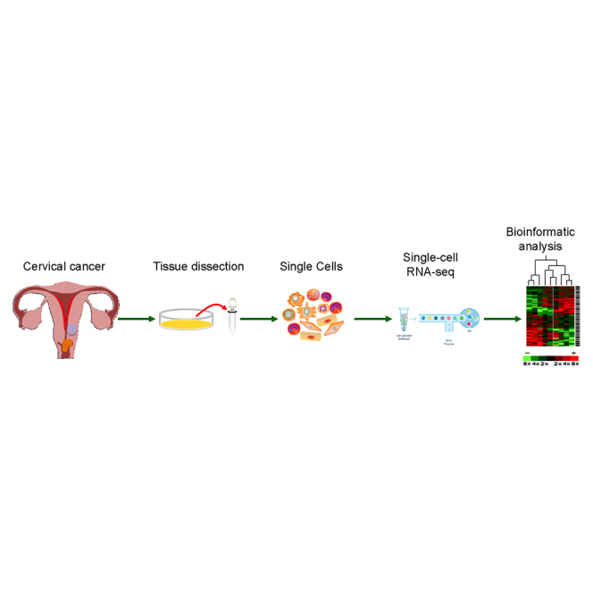
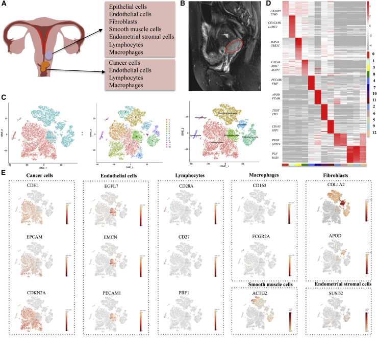
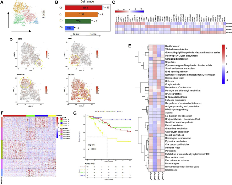
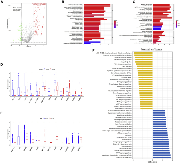
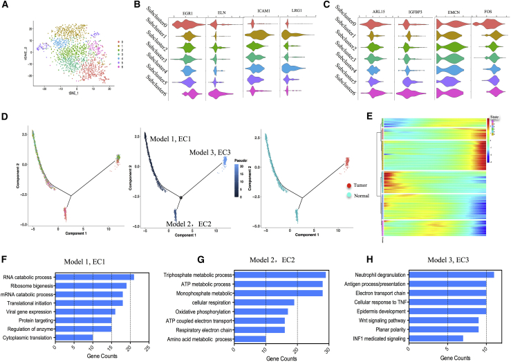
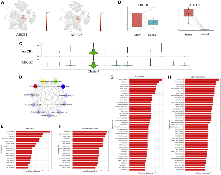
Cervical cancer (CC) is the fourth leading cause of deaths in gynecological malignancies. Although the etiology of CC has been extensively investigated, the exact pathogenesis of CC remains incomplete. Recently, single-cell technologies demonstrated advantages in exploring intra-tumoral diversification among various tumor cells. However, single-cell transcriptome analysis (single-cell RNA sequencing [scRNA-seq]) of CC cells and microenvironment has not been conducted. In this study, a total of 20,938 cells from CC and adjacent normal tissues were examined by scRNA-seq. We identified four tumor cell subpopulations in tumor cells, which had specific signature genes with different biological functions and presented different prognoses. Among them, we identified a subset of cancer stem cells (CSCs) that was related to the developmental hierarchy of tumor progression. Then, we compared the expressive differences between tumor-derived endothelial cells (TECs) and normal ECs (NECs) and revealed higher expression of several metabolism-related genes in TECs. Then, we explored the potential biological function of ECs in vascularization and found several marker genes, which played a prior role in connections between cancer cells and ECs. Our findings provide valuable resources for deciphering the intra-tumoral heterogeneity of CC and uncover the developmental procedure of ECs, which paves the way for CC therapy.
Keywords: cervical cancer; endothelial cell; single-cell RNA sequencing; tumor heterogeneity.
© 2021 The Authors.
Conflict of interest statement
The authors declare no competing interests.
Figures

Similar articles
-
Single-cell transcriptomics reveals cellular heterogeneity and molecular stratification of cervical cancer.Commun Biol. 2022 Nov 10;5(1):1208. doi: 10.1038/s42003-022-04142-w. Commun Biol. 2022. PMID: 36357663 Free PMC article.
-
Large-Scale Single-Cell RNA-Seq Reveals Molecular Signatures of Heterogeneous Populations of Human Induced Pluripotent Stem Cell-Derived Endothelial Cells.Circ Res. 2018 Aug 3;123(4):443-450. doi: 10.1161/CIRCRESAHA.118.312913. Circ Res. 2018. PMID: 29986945 Free PMC article.
-
Single-cell transcriptome profiling reveals vascular endothelial cell heterogeneity in human skin.Theranostics. 2021 Apr 19;11(13):6461-6476. doi: 10.7150/thno.54917. eCollection 2021. Theranostics. 2021. PMID: 33995668 Free PMC article.
-
Cellular heterogeneity and stem cells of vascular endothelial cells in blood vessel formation and homeostasis: Insights from single-cell RNA sequencing.Front Cell Dev Biol. 2023 Mar 21;11:1146399. doi: 10.3389/fcell.2023.1146399. eCollection 2023. Front Cell Dev Biol. 2023. PMID: 37025170 Free PMC article. Review.
-
Deciphering cell-cell interactions and communication in the tumor microenvironment and unraveling intratumoral genetic heterogeneity via single-cell genomic sequencing.Bioengineered. 2022 Jul-Dec;13(7-12):14974-14986. doi: 10.1080/21655979.2023.2185434. Bioengineered. 2022. PMID: 37105769 Free PMC article. Review.
Cited by
-
A risk prediction model mediated by genes of APOD/APOC1/SQLE associates with prognosis in cervical cancer.BMC Womens Health. 2022 Dec 19;22(1):534. doi: 10.1186/s12905-022-02083-4. BMC Womens Health. 2022. PMID: 36536343 Free PMC article.
-
Cervical cancer immune infiltration microenvironment identification, construction of immune scores, assisting patient prognosis and immunotherapy.Front Immunol. 2023 Mar 10;14:1135657. doi: 10.3389/fimmu.2023.1135657. eCollection 2023. Front Immunol. 2023. PMID: 36969161 Free PMC article.
-
Targeting tumor-associated macrophage-derived CD74 improves efficacy of neoadjuvant chemotherapy in combination with PD-1 blockade for cervical cancer.J Immunother Cancer. 2024 Aug 6;12(8):e009024. doi: 10.1136/jitc-2024-009024. J Immunother Cancer. 2024. PMID: 39107132 Free PMC article.
-
Comparative profiling of single-cell transcriptome reveals heterogeneity of tumor microenvironment between solid and acinar lung adenocarcinoma.J Transl Med. 2022 Sep 23;20(1):423. doi: 10.1186/s12967-022-03620-3. J Transl Med. 2022. PMID: 36138435 Free PMC article.
-
Identification of Siglec-10 as a new dendritic cell checkpoint for cervical cancer immunotherapy.J Immunother Cancer. 2024 Aug 28;12(8):e009404. doi: 10.1136/jitc-2024-009404. J Immunother Cancer. 2024. PMID: 39209455 Free PMC article.
References
LinkOut - more resources
Full Text Sources
Other Literature Sources
