. 2021 Apr;18(2):1273-1294.
doi: 10.1007/s13311-020-00999-z. Epub 2021 Jan 21.

Decreased Level of Exosomal miR-5121 Released from Microglia Suppresses Neurite Outgrowth and Synapse Recovery of Neurons Following Traumatic Brain Injury

Affiliations

Decreased Level of Exosomal miR-5121 Released from Microglia Suppresses Neurite Outgrowth and Synapse Recovery of Neurons Following Traumatic Brain Injury

Chengcheng Zhao et al. Neurotherapeutics. 2021 Apr.

Abstract

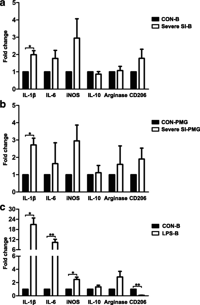
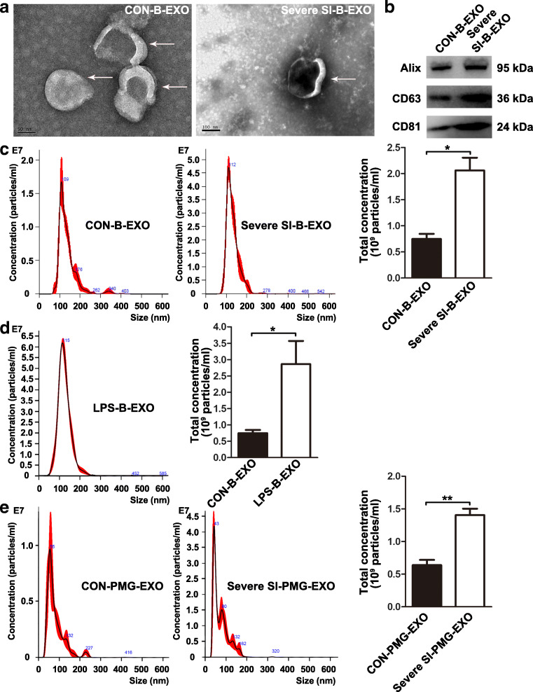
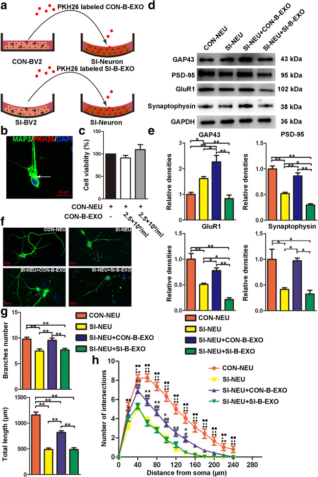
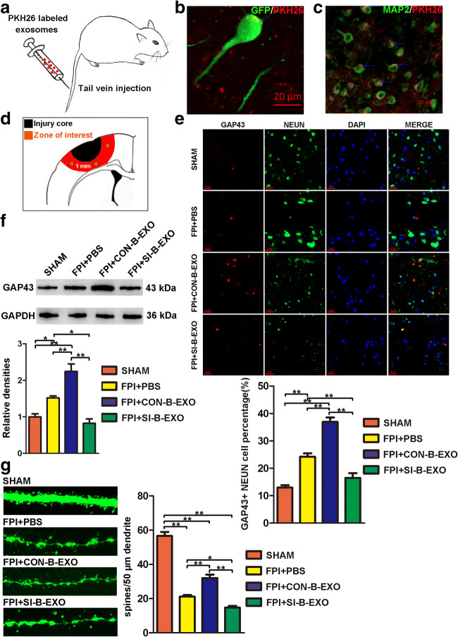
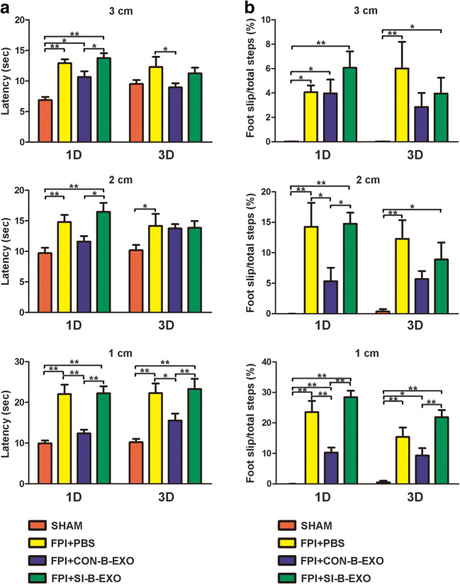
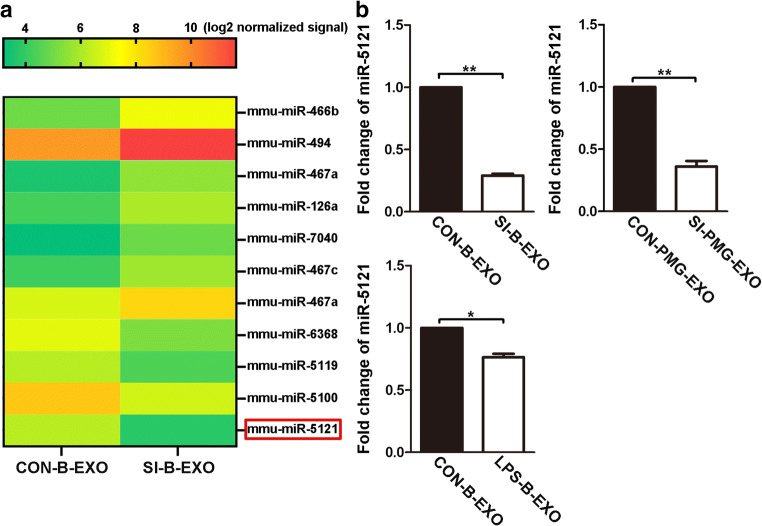
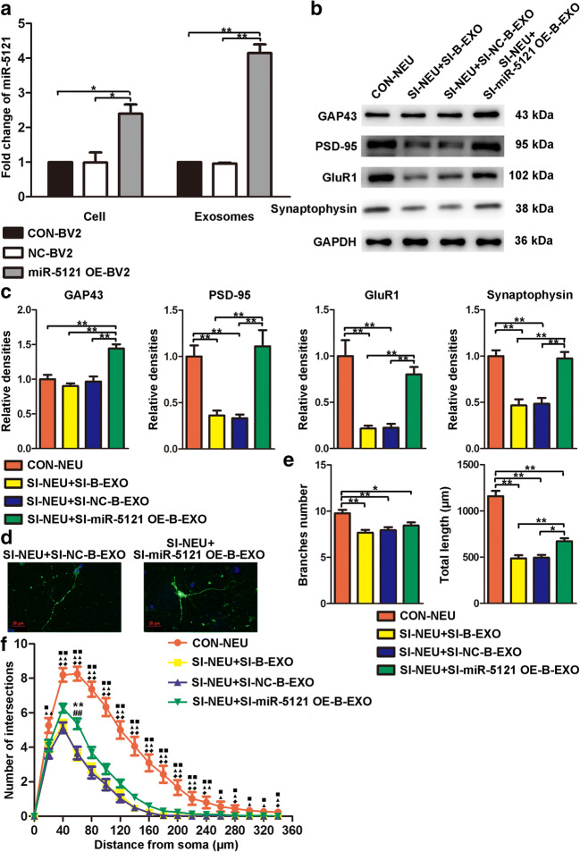
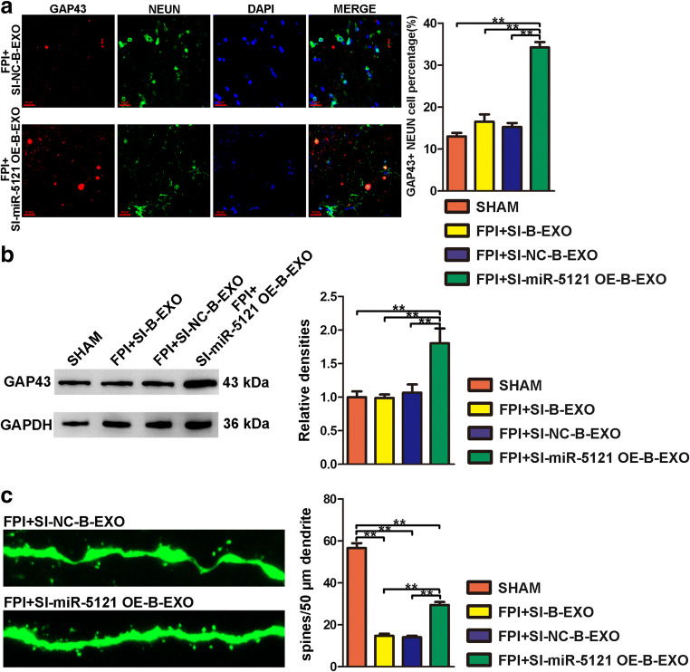
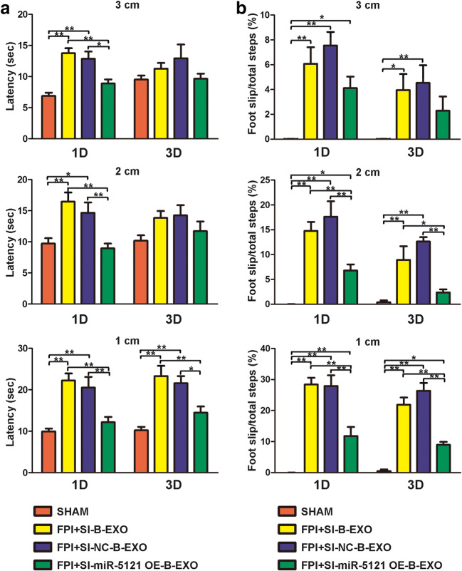
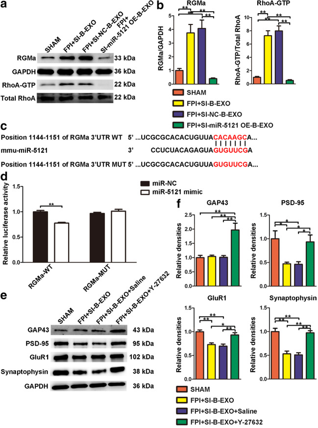
Activated microglia can suppress neurite outgrowth and synapse recovery in the acute stage following traumatic brain injury (TBI). However, the underlying mechanism has not been clearly elucidated. Exosomes derived from microglia have been reported to play a critical role in microglia-neuron interaction in healthy and pathological brains. Here, we aimed to investigate the role of microglia-derived exosomes in regulating neurite outgrowth and synapse recovery following TBI. In our study, exosomes derived from microglia were co-cultured with stretch-injured neurons in vitro and intravenously injected into mice that underwent fluid percussion injury (FPI) by tail vein injection in vivo. The results showed that microglia-derived exosomes could be absorbed by neurons in vitro and in vivo. Moreover, exosomes derived from stretch-injured microglia decreased the protein levels of GAP43, PSD-95, GluR1, and Synaptophysin and dendritic complexity in stretch-injured neurons in vitro, and reduced GAP43+ NEUN cell percentage and apical dendritic spine density in the pericontusion region in vivo. Motor coordination was also impaired in mice treated with stretch-injured microglia-derived exosomes after FPI. A microRNA microarray showed that the level of miR-5121 was decreased most greatly in exosomes derived from stretch-injured microglia. Overexpression of miR-5121 in stretch-injured microglia-derived exosomes partly reversed the suppression of neurite outgrowth and synapse recovery of neurons both in vitro and in vivo. Moreover, motor coordination in miR-5121 overexpressed exosomes treated mice was significantly improved after FPI. Following mechanistic study demonstrated that miR-5121 might promote neurite outgrowth and synapse recovery by directly targeting RGMa. In conclusion, our finding revealed a novel exosome-mediated mechanism of microglia-neuron interaction that suppressed neurite outgrowth and synapse recovery of neurons following TBI.

Keywords: Exosomes; microRNA; microglia; neuron; traumatic brain injury.


Conflict of interest statement

The authors declare that they have no conflict of interest.

Figures

Fig. 1
Fig. 1
(A) IL-1β, IL-6, iNOS, IL-10, Arginase, and CD206 mRNA expression of BV2 microglia at 24 h after severe stretch-induced injury. (B) IL-1β, IL-6, iNOS, IL-10, Arginase, and CD206 mRNA expression of primary microglia at 24 h after severe stretch-induced injury. (C) IL-1β, IL-6, iNOS, IL-10, Arginase, and CD206 mRNA expression of BV2 microglia at 24 h after LPS stimulation. Quantification revealed that the expression level of IL-1β mRNA was significantly increased in BV2 microglia and primary microglia after severe stretch-induced injury. The expression levels of IL-1β, IL-6, and iNOS mRNA were significantly increased and the expression level of CD206 was significantly decreased in BV2 microglia at 24 h after LPS stimulation. All data are represented as the mean ± SEM; n = 3; two-tailed t tests was used to analyze significant differences. *p < 0.05, **p < 0.01
Fig. 2
Fig. 2
Characterization of exosomes derived from microglia. (A) The structure of uninjured BV2 and stretch-injured BV2-derived exosomes observed by TEM. (B) Western blot analysis of exosomal surface markers Alix, CD63, and CD81 in BV2-derived exosomes. (C) Particle size distribution and total concentration of uninjured BV2 and stretch-injured BV2-derived exosomes measured by nanoparticle tracking analysis. (D) Particle size distribution and total concentration of LPS-stimulated BV2-derived exosomes measured by nanoparticle tracking analysis. (E) Particle size distribution and total concentration of uninjured primary microglia and stretch-injured primary microglia-derived exosomes measured by nanoparticle tracking analysis. Quantification revealed that the total concentration of microglia-derived exosomes was significantly increased in the Severe SI-B-EXO group and LPS-B-EXO group compared to that of the CON-B-EXO group. The total concentration of microglia-derived exosomes was significantly increased in the Severe SI-PMG-EXO group compared to that of the CON-PMG-EXO group. All data are represented as the mean ± SEM; n = 3; two-tailed t tests was used to analyze significant differences. *p < 0.05, **p < 0.01
Fig. 3
Fig. 3
Exosomes derived from stretch-injured BV2 microglia suppressed neurite outgrowth and synapse recovery in vitro. (A) A schematic diagram illustrating fluorescent labeling of uninjured BV2 and stretch-injured BV2-derived exosomes with PKH26 and co-culture of PKH26-labeled exosomes with stretch-injured neurons. (B) Confocal imaging showed that PKH26 (red)-labeled exosomes derived from BV2 microglia were taken up by neurons in vitro. Neurons were identified by MAP2 (green). (C) Cell viability of neurons co-cultured with two concentrations of uninjured BV2-derived exosomes (2.5 × 108/ml and 2.5 × 109/ml) was detected by CCK-8 assay. Quantification revealed that uninjured BV2-derived exosomes (2.5 × 108/ml and 2.5 × 109/ml) showed no significant cytotoxicity against neurons after 24 h (n = 3). (D) Representative immunoblots of GAP43, PSD-95, GluR1, and Synaptophysin in the CON-NEU group, SI-NEU group, SI-NEU+CON-B-EXO group, and SI-NEU+SI-B-EXO group. (E) Quantification revealed that the protein levels of GAP43, PSD-95, GluR1, and Synaptophysin were decreased in stretch-injured neurons co-cultured with exosomes derived from stretch-injured BV2 microglia compared with that of exosomes derived from uninjured BV2 microglia. (F) Representative confocal imaging of uninjured neurons, stretch-injured neurons, and stretch-injured neurons co-cultured with uninjured BV2 and stretch-injured BV2-derived exosomes. Neurons were identified by MAP2 (green). (G) Quantified results of neurite analysis revealed that branch number and total length were significantly decreased in stretch-injured neurons co-cultured with exosomes derived from stretch-injured BV2 microglia compared with that of exosomes derived from uninjured BV2 microglia. (H) Sholl analysis identified a decrease in the number of intersections 40, 60, 80, 100, and 140 μm from the neuron soma in the SI-NEU+SI-B-EXO group compared with that of the SI-NEU+CON-B-EXO group. Two black squares, p < 0.01, CON-NEU versus SI-NEU; one black triangle, p < 0.05, two black triangles, p < 0.01, CON-NEU versus SI-NEU+CON-B-EXO; two black diamonds, p < 0.01, CON-NEU versus SI-NEU+SI-B-EXO; *p < 0.05, **p < 0.01, SI-NEU versus SI-NEU+CON-B-EXO; #p < 0.05, ##p < 0.01, SI-NEU+CON-B-EXO versus SI-NEU+SI-B-EXO. All data are represented as the mean ± SEM; n = 6; one-way ANOVA with Tukey’s post hoc test was used to analyze significant differences. *p < 0.05, **p < 0.01
Fig. 4
Fig. 4
Tail vein injection of exosomes derived from stretch-injured BV2 microglia suppressed neurite outgrowth and synapse recovery in vivo. (A) A schematic diagram illustrating tail vein injection of PKH26-labeled exosomes. (B) Representative confocal imaging of PKH26-labeled exosomes absorbed by GFP+ pyramid neurons in the pericontusion region. (C) Representative confocal imaging of PKH26-labeled exosomes absorbed by GFP- neurons in the pericontusion region. Neurons were identified by MAP2 (green). (D) A schematic diagram of regions of interest (within 1 mm from the edge of injury core). Images were taken at both sides and the bottom of regions of interest (squares). (E) Detection of GAP43 expression in neurons by immunofluorescence. Quantification revealed that tail vein injection of exosomes derived from stretch-injured BV2 microglia decreased the numbers of GAP43+ neurons. Neurons were identified by NEUN (green). (F) Representative immunoblots of GAP43 in the SHAM group, FPI+PBS group, FPI+CON-B-EXO group, and FPI+SI-B-EXO group. Quantification revealed that tail vein injection of exosomes derived from stretch-injured BV2 microglia significantly decreased the protein level of GAP43 in the pericontusion region. (G) Representative confocal imaging of apical dendritic spines in the pericontusion region (layers V/VI). Quantification revealed that tail vein injection of exosomes derived from stretch-injured BV2 microglia decreased apical dendritic spine density in the pericontusion region after FPI. All data are represented as the mean ± SEM; n = 6; one-way ANOVA with Tukey’s post hoc test was used to analyze significant differences. *p < 0.05, **p < 0.01
Fig. 5
Fig. 5
Tail vein injection of exosomes derived from stretch-injured BV2 microglia impaired motor coordination in mice after FPI. (A) Latency and (B) percentage of foot-slips in the beam walking test at 1 and 3 days following FPI were measured. Quantification revealed that SI-B-EXO-treated mice performed a significant increase in time and percentage of foot-slips when they crossed the 2-cm and 1-cm width beams in comparison to CON-B-EXO-treated mice at 1 day after FPI. For the 3-cm width beam, SI-B-EXO-treated mice only performed a significant increase in time to cross the beam and showed no significant difference in percentage of foot-slips compared with CON-B-EXO-treated mice. At 3 days after FPI, SI-B-EXO-treated mice performed a significant increase in time and percentage of foot-slips when they crossed the 1-cm width beam in comparison to CON-B-EXO-treated mice. There was no significant difference between SI-B-EXO-treated and CON-B-EXO-treated mice in time and percentage of foot-slips to cross the 3-cm and 2-cm width beams. Moreover, mice in the FPI+SI-B-EXO group showed no significant difference compared to mice in the FPI+PBS group in latency and percentage of foot-slips at both 1 and 3 days following FPI. All data are represented as the mean ± SEM; n = 7; two-way repeated measures ANOVA with Bonferroni post hoc test was used to analyze significant differences. *p < 0.05, **p < 0.01
Fig. 6
Fig. 6
The level of miR-5121 was decreased in exosomes derived from stretch-injured microglia and LPS-stimulated microglia. (A) Heat map of microRNA expression normalized data of exosomes derived from control and injured BV2 microglia detected by microarray analysis. MicroRNAs with fold change > 3 were displayed in heat map. (B) The miR-5121 level in exosomes derived from microglia was detected by quantitative real-time PCR. Quantification revealed that the level of miR-5121 were decreased in exosomes derived from stretch-injured BV2 microglia, stretch-injured primary microglia, and LPS-stimulated microglia. All data are represented as the mean ± SEM; n = 3; two-tailed t tests was used to analyze significant differences. *p < 0.05, **p < 0.01
Fig. 7
Fig. 7
MiR-5121 overexpressed exosomes derived from stretch-injured BV2 microglia promoted neurite outgrowth and synapse recovery in vitro. (A) The miR-5121 levels in BV2 microglia and exosomes derived from BV2 microglia were detected by quantitative real-time PCR. Quantification revealed that the levels of miR-5121 were elevated both in BV2 microglia and in exosomes derived from BV2 microglia in the miR-5121 OE-BV2 group (n = 3). (B) Representative immunoblots of GAP43, PSD-95, GluR1, and Synaptophysin in the CON-NEU group, SI-NEU+SI-B-EXO group, SI-NEU+SI-NC-B-EXO group, and SI-NEU+SI-miR-5121 OE-B-EXO group. (C) Quantification revealed that the protein levels of GAP43, PSD-95, GluR1, and Synaptophysin were significantly increased in stretch-injured neurons co-cultured with miR-5121 overexpressed exosomes derived from stretch-injured BV2 microglia. (D) Representative confocal imaging of stretch-injured neurons co-cultured with exosomes derived from stretch-injured negative control BV2 and stretch-injured miR-5121 overexpressed BV2. Neurons were identified by MAP2 (green). (E) Quantified results of neurite analysis revealed that total length was significantly increased in stretch-injured neurons co-cultured with miR-5121 overexpressed exosomes derived from stretch-injured BV2 microglia. (F) Sholl analysis identified an increase in the number of intersections 60 μm from the neuron soma in the SI-NEU+SI-miR-5121 OE-B-EXO group compared with that of the SI-NEU+SI-B-EXO group and SI-NEU+SI-NC-B-EXO group. One black square, p < 0.05; two black squares, p < 0.01, CON-NEU versus SI-NEU+SI-B-EXO; one black triangle, p < 0.05; two black triangles, p < 0.01, CON-NEU versus SI-NEU+SI-NC-B-EXO; one black diamond, p < 0.05; two black diamonds, p < 0.01, CON-NEU versus SI-NEU+SI-miR-5121 OE-B-EXO; **p < 0.01, SI-NEU+SI-B-EXO versus SI-NEU+SI-miR-5121 OE-B-EXO; ##p < 0.01, SI-NEU+SI-NC-B-EXO versus SI-NEU+SI-miR-5121 OE-B-EXO. All data are represented as the mean ± SEM; n = 6; one-way ANOVA with Tukey’s post hoc test was used to analyze significant differences. *p < 0.05, **p < 0.01
Fig. 8
Fig. 8
Tail vein injection of miR-5121 overexpressed exosomes derived from stretch-injured BV2 microglia promoted neurite outgrowth and synapse recovery in vivo. (A) Detection of GAP43 expression in neurons by immunofluorescence. Quantification revealed that tail vein injection of miR-5121 overexpressed exosomes derived from stretch-injured BV2 microglia increased the numbers of GAP43+ neurons. Neurons were identified by NEUN (green). (B) Representative immunoblots of GAP43 in the SHAM group, FPI+SI-B-EXO group, FPI+SI-NC-B-EXO group, and FPI+SI-miR-5121 OE-B-EXO group. Quantification revealed that tail vein injection of miR-5121 overexpressed exosomes derived from stretch-injured BV2 microglia significantly increased the protein level of GAP43 in the pericontusion region. (C) Representative confocal imaging of apical dendritic spines in the pericontusion region (layers V/VI). Quantification revealed that tail vein injection of miR-5121 overexpressed exosomes derived from stretch-injured BV2 microglia increased apical dendritic spine density in the pericontusion region after FPI. All data are represented as the mean ± SEM; n = 6; one-way ANOVA with Tukey’s post hoc test was used to analyze significant differences. **p < 0.01
Fig. 9
Fig. 9
Tail vein injection of miR-5121 overexpressed exosomes derived from stretch-injured BV2 microglia improved motor coordination in mice after FPI. (A) Latency and (B) percentage of foot-slips in the beam walking test at 1 and 3 days following FPI were measured. Quantification revealed that mice in the FPI+SI-miR-5121 OE-B-EXO group crossed the beams significantly faster than mice in the FPI+SI-B-EXO group and FPI+SI-NC-B-EXO group in both 3-cm, 2-cm, and 1-cm width trials at 1 day following FPI and only in the 1-cm width trial at 3 days following FPI. Mice in the FPI+SI-miR-5121 OE-B-EXO group showed significant decreases in the percentage of foot-slips compared with mice in the FPI+SI-B-EXO group and FPI+SI-NC-B-EXO group in 2-cm and 1-cm width trials at both 1 day and 3 days following FPI. All data are represented as the mean ± SEM; n = 7; two-way repeated measures ANOVA with Bonferroni post hoc test was used to analyze significant differences. *p < 0.05, **p < 0.01
Fig. 10
Fig. 10
Exosomal miR-5121 targeted 3′UTR of RGMa. (A) Representative immunoblots of RGMa, RhoA-GTP, and total RhoA in the SHAM group, FPI+SI-B-EXO group, FPI+SI-NC-B-EXO group, and FPI+SI-miR-5121 OE-B-EXO group. (B) Quantification revealed that the expression levels of RGMa and RhoA-GTP was significantly decreased in the FPI+SI-miR-5121 OE-B-EXO group compared with that of the FPI+SI-B-EXO group and FPI+SI-NC-B-EXO group. (C) Wild-type and mutation-type of the RGMa 3′UTR binding site of miR-5121. (D) Quantification revealed that miR-5121 mimic reduced the luciferase activity in 293T cells transfected with the pmirGLO-RGMa-3′UTR-WT reporter, but not in 293T cells transfected with the pmirGLO-RGMa-3′UTR-MUT reporter (n = 3). (E) Representative immunoblots of GAP43, PSD-95, GluR1, and Synaptophysin in the SHAM group, FPI+SI-B-EXO group, FPI+SI-B-EXO+Saline group, and FPI+SI-B-EXO+Y-27632 group. (F) Quantification revealed that Rho kinase inhibitor, Y-27632, significantly increased the protein levels of GAP43, PSD-95, GluR1, and Synaptophysin in the pericontusion region. All data are represented as the mean ± SEM; n = 6; one-way ANOVA with Tukey’s post hoc test and two-tailed t tests were used to analyze significant differences. *p < 0.05, **p < 0.01

Similar articles

Cited by

References

    1. Popescu C, Anghelescu A, Daia C, Onose G. Actual data on epidemiological evolution and prevention endeavours regarding traumatic brain injury. Journal of medicine and life. 2015;8(3):272–277. - PMC - PubMed
    1. Jiang JY, Gao GY, Feng JF, et al. Traumatic brain injury in China. The Lancet Neurology. 2019;18(3):286–295. doi: 10.1016/S1474-4422(18)30469-1. - DOI - PubMed
    1. Rosenfeld JV, Maas AI, Bragge P, Morganti-Kossmann MC, Manley GT, Gruen RL. Early management of severe traumatic brain injury. Lancet. 2012;380(9847):1088–1098. doi: 10.1016/S0140-6736(12)60864-2. - DOI - PubMed
    1. Gruenbaum SE, Zlotnik A, Gruenbaum BF, Hersey D, Bilotta F. Pharmacologic Neuroprotection for Functional Outcomes After Traumatic Brain Injury: A Systematic Review of the Clinical Literature. CNS drugs. 2016;30(9):791–806. doi: 10.1007/s40263-016-0355-2. - DOI - PMC - PubMed
    1. Tsitsopoulos PP, Abu Hamdeh S, Marklund N. Current Opportunities for Clinical Monitoring of Axonal Pathology in Traumatic Brain Injury. Frontiers in neurology. 2017;8:599. doi: 10.3389/fneur.2017.00599. - DOI - PMC - PubMed

Publication types