. 2021 Jan 6;40(1):9.
doi: 10.1186/s13046-020-01819-0.

Lnc-GAN1 expression is associated with good survival and suppresses tumor progression by sponging mir-26a-5p to activate PTEN signaling in non-small cell lung cancer

Affiliations

Lnc-GAN1 expression is associated with good survival and suppresses tumor progression by sponging mir-26a-5p to activate PTEN signaling in non-small cell lung cancer

Rui-Qi Wang et al. J Exp Clin Cancer Res. .

Abstract

Background: Long non-coding RNAs (lncRNAs) play vital roles in the development and progression of non-small-cell lung cancer (NSCLC); however, the role of most lncRNAs in NSCLC remains unknown. This study explored the clinical significance, biological function and underlying mechanism of lnc-GAN1 in NSCLC.

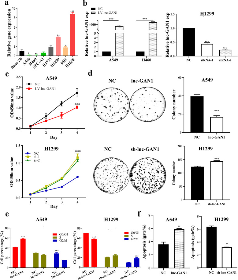
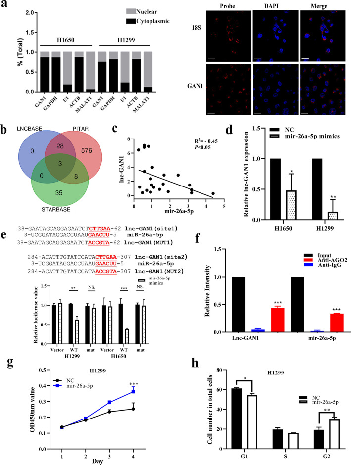
Methods: With a custom lncRNA microarray we found that lnc-GAN1 is markedly downregulated in NSCLC tissues. Then lnc-GAN1 expression level was measured using qRT-PCR in NSCLC tissues and cell lines. Survival was assessed using the Kaplan-Meier method. The biological functions of lnc-GAN1 in lung cancer cells were evaluated in vitro and in vivo. RNA fluorescence in situ hybridization and subcellular localization assays revealed the subcellular distribution of lnc-GAN1 in cells. Bioinformatic analysis was adopted to predict miRNAs and signaling pathways regulated by lnc-GAN1. RNA immunoprecipitation and Dual-luciferase reporter assays were used to assess the interaction between lnc-GAN1 and miR-26a-5p in lung cancer cells.

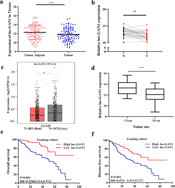
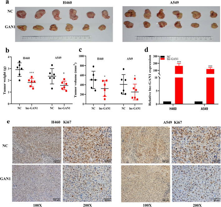
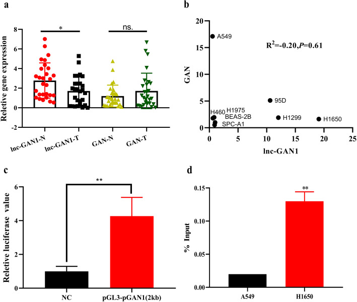
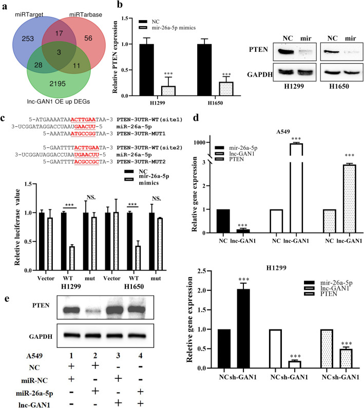
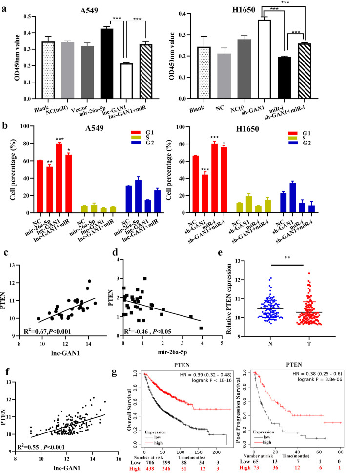
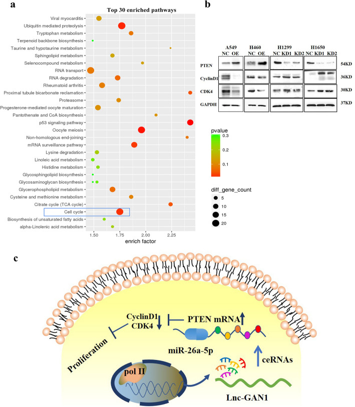
Results: lnc-GAN1 is downregulated in HCC tissues and associated with larger tumor size and poor overall survival and disease-free survival; its ectopic expression suppresses cell proliferation, colony formation, and cell cycle progression and induces apoptosis in NSCLC cells; it also inhibits tumor growth in the NSCLC xenograft model. We further proved that lnc-GAN1 is localized in cytoplasm and transcribed independently from its parental gene GAN. Mechanistically, lnc-GAN1 acts as a sponge for miR-26a-5p by two seed sequences, and the two non-coding RNAs have a negative relationship in NSCLC tissues; we further prove that PTEN is a direct target of miR-26a-5p and lnc-GAN1 inhibits cell cycle signaling pathway by activating PTEN, whose expression level correlated negatively with miR-26a-5p level but positively with lnc-GAN1 level in NSCLC samples.

Conclusions: Lnc-GAN1 is downregulated and associated with poor survival of NSCLC patients, and mechanistically acts as a tumor suppressor via sponging and inhibiting miR-26a-5p to upregulate PTEN. This study provides a potential prognostic biomarker and treatment target for NSCLC.

Keywords: Lnc-GAN1; Lung cancer; PTEN; miR-26a-5p.


Conflict of interest statement

The authors declare that they have no competing interests.

Figures

Fig. 1
Fig. 1
Lnc-GAN1 is downregulated in NSCLC tissues. a Lnc-GAN1 is significantly downregulated in 194 NSCLC tissues compared with 100 matched adjacent normal lung tissue detected by lncRNA microarray (*** P < 0.001). b lnc-GAN1 levels in 30 pairs NSCLC and adjacent tissues were measured by real-time qRT-PCR. lnc-GAN1 level is significantly lower in NSCLC tissues than in the paired adjacent normal lung tissues. (* P < 0.05). c lnc-GAN1 expression level is lower in lung adenocarcinomas (n = 483) than in the normal lung tissues (n = 347) obtained from TCGA (* P < 0.05). d lnc-GAN1 is highly expressed in NSCLC tumors less than 3 cm than in those larger than or equal to 3 cm (*** P < 0.001). e, f Kaplan-Meier survival analysis suggests that NSCLC patients with high lnc-GAN1 levels had better overall survival (OS) and disease-free survival (DFS) than those with low lnc-GAN1 expression
Fig. 2
Fig. 2
Lnc-GAN1 functions as a tumor suppressor for NSCLC cells in vitro. a Lnc-GAN1 levels were measured by real-time qRT-PCR in the NSCLC cell lines A549, H-460, H1299, H1650, SPC-A1, H1975, 95D, and in a human lung epithelial cell line BEAS-2B. b The histograms represent Lnc-GAN1 expression levels detected by qRT-PCR in A549 and H460 cells with overexpression of lnc-GAN1 or control vector, and in H1299 transfected with siRNA against lnc-GAN1 or control siRNA. c Proliferation curves of A549 cells with overexpression of lnc-GAN1 or control vector and H1299 cells transfected with sh-lnc-GAN1 or control shRNA, as determined by CCK8 assay. The results show that overexpressed lnc-GAN1 represses cell proliferation in A549 cells and sh-lnc-GAN1 has the reverse effects in H1299 cells. d Colony formation assay was performed to evaluate the oncogenic growth of A549 cells overexpressing lnc-GAN1 or control vector and H1299 cells transfected with sh-lnc-GAN1 or control. The right histograms denote the colony number of and of the A549 and H1299 cells, respectively, indicating that overexpressed lnc-GAN1 inhibits colony formation of A549 cells and sh-lnc-GAN1 enhances colony formation of H1299 cells. e The histograms designate percentage changes of G0/G1, S, and G2/M phases in A549 cells overexpressing lnc-GAN1 or control vector and H1299 cells transfected with sh-lnc-GAN1 or control, as detected by flow cytometry assay. The result demonstrates that overexpressed lnc-GAN1 induces cell cycle arrest in G0/G1 phase of A549 cells, and silencing lnc-GAN1 has the reverse effects on H1299. f The histograms represent the apoptosis percentage of A549 overexpressing lnc-GAN1 or control vector and H1299 cells transfected with sh-lnc-GAN1 or control shRNA, as detected by flow cytometry assay. The result suggests that overexpressed lnc-GAN1 induces apoptosis in A549 cells, and silencing lnc-GAN1 has the reverse effects on H1299 cells. Data represent mean ± SD of three independent experiments (*P < 0.05; ***P < 0.001, by Student’s t-test)
Fig. 3
Fig. 3
Lnc-GAN1 represses NSCLC cell growth in vivo. a The tumor masses are derived from H460 and A549 cells with overexpression of lnc-GAN1 or control vector, which were subcutaneously inoculated into the flank region of 6 nude mice, respectively. Upper penal: Negative controls; lower penal: lnc-GAN1 overexpression. b, c The weights (b) and volume (c) of the tumors derived from H460 and A549 cells with overexpression of Lnc-GAN1 or control vector, respectively. d Lnc-GAN1 extracted from  Xenotransplanted tumors was detected by qRT-PCR. e Ki-67 protein levels were detected with IHC in tumors derived from A549 or H460 cells with overexpressing lnc-GAN1 or control vector. *P < 0.05; ***P < 0.001
Fig. 4
Fig. 4
Lnc-GAN1 is expressed independent of its parent gene GAN. a Lnc-GAN1 expression is lower in 30 NSCLC tissues than in paired normal lung tissues, while its parent gene GAN expression is not different between the same NSCLC and paired normal lung tissues. b There is not a correlation between Lnc-GAN1 and GAN expressions in 8 cell lines. c In dual-luciferase reporter assay, A549 cells with the reporter containing 2000-bp DNA sequence upstream of lnc-GAN1 produce significantly higher Luciferase activity than A549 cells with control reporter, indicating that the 2000-bp sequence harbors promoter binding site. Luciferase activity was calculated as a firefly/renilla luciferase ratio and expressed as mean ± SD of three independent experiments. d Chromatin immunoprecipitation (ChIP) analysis on H460 and H1650 cells with Pol II antibody and the precipitated DNAs were amplified by PCR with the primers for the 2000-bp DNA sequence upstream of lnc-GAN1. H1650 cells with high expression of lnc-GAN1 show remarkably higher PCR product than H460 cells with very low expression of lnc-GAN1, suggesting that this DNA sequence has high Pol II activity in H1650 cells
Fig. 5
Fig. 5
Lnc-GAN1 located in cytoplasm sponges and inhibits miR-26a-5p. a Left-Panel: Lnc-GAN1 is mainly located in the cytoplasm of H1299 and H1650 cells, as measured by real-time qPCR on cytoplasm and nucleus, respectively. GAPDH mRNA was used as a cytoplasmic control, while U1 snRNA was used as a nuclear control. Right-Panel: lnc-GAN1 was visually observed in the cytoplasm of H460 cells, as shown by FISH. 18S was used as the positive control in cytoplasm. b Venn diagram shows 3 shared predicted targets (miRNAs) of lnc-GAN1 by three databases, including miR-26a-5p, mir-26b-5p and mir-1297. c There is a significantly negative correlation between lnc-GAN1 and mir-26a-5p levels in 30 NSCLC samples, as determined by qRT-PCR (R2= − 0.45, P < 0.05). d Ectopic miR-26a-5p expression significantly reduced lnc-GAN1 mRNA levels in H1299 and H1650 cells (* P < 0.05), as measured by qRT-PCR. e Upper-Panel: The predicted wild-type seed sequences of lnc-GAN1 complementary with miR-26a-5p and investigator-designed mutated seed sequences. Lower-panel: Histogram shows that when miR-26a-5p was overexpressed in H1299 or H1650 cells, the luciferase activity of cells with wild-type seed sequence of lnc-GAN1 was sharply reduced compared with that of control cells, but not changed in cells with mutant miR-26a-5p binding sites, as assayed by Dual-luciferase reporter assays, indicating that there are direct binding sites between lnc-GAN1 and miR-26a-5p. f RIP was performed on H1650 cell lysates with mouse anti-AGO2 or IgG. The precipitated lnc-GAN1 and miR-26a-5p were evaluated by real-time qRT-PCR, and both lnc-GAN1 and miR-26a-5p were enriched in the same precipitation by anti-AGO2 (*** P < 0.001). g Overexpressed miR-26a-5p promoted proliferation of H1299 cells, as shown by CCK8 assay. h Overexpressed miR-26a-5p accelerated cell cycle progression in H1299 cells. Data represent the mean ± SD of three independent experiments
Fig. 6
Fig. 6
Lnc-GAN1 upregulates PTEN by inhibiting miR-26a-5p. a Venn diagram shows 3 shared predicted target genes of miR-26a-5p by two miRNA databases and overlapped with upregulated ones induced by expressed lnc-GAN1, including PTEN, CDK8, and PTGS2. b MiR-26a-5p overexpression reduces PTEN mRNA and protein levels in H1299 and H1650 cells (***P < 0.001), as determined by qRT-PCR and western blot. c Upper-Panel: The predicted wild-type seed sequences of PTEN 3’UTR complementary with miR-26a-5p and investigator-designed mutated seed sequences. Lower-panel: Histogram shows that when miR-26a-5p is overexpressed in H1299 and H1650 cells, the luciferase activity of the cells with wild-type seed sequence of PTEN 3’UTR is markedly reduced compared with that of control cells, but not changed in the cells with mutant miR-26a-5p binding sites, as assayed by Dual-luciferase reporter assays. d Lnc-GAN1 overexpression significantly decreases miR-26a-5p levels and increases PTEN mRNA levels in A549 cells, whereas lnc-GAN1 knockdown exerts the opposite effects in H1299 cells. e PTEN protein level is downregulated in A549 cells with miR-26a-5p overexpression, upregulated in the cells with lnc-GAN1 expression and not changed in the cells co-transfected with lnc-GAN1 and miR-26a-5p
Fig. 7
Fig. 7
lnc-GAN1 inhibits NSCLC progression by repressing miR-26a-5p to activate PTEN signaling. a miR-26a-5p overexpression alone markedly increases the cell proliferation and lnc-GAN1 overexpression alone remarkably decreases cell proliferation in A549 cells compared with blank or respective control treatments, whereas when both are overexpressed, cell proliferation is not changed; downregulation of miR-26a-5p alone or lnc-GAN1 alone or both combination produces opposite effects on cell proliferation in H1650 cells. b miR-26a-5p overexpression alone significantly promotes cell cycle progression and lnc-GAN1 overexpression alone has the reverse effect on cell cycle, whereas when both are overexpressed, the cell cycle is not changed; downregulation of miR-26a-5p alone or lnc-GAN1 alone or both combination produces opposite effects on cell cycle in H1650 cells. c There is a positive correlation between lnc-GAN1 and PTEN levels in 30 NSCLC tissues, as measured by qRT-PCR (R2 = 0.55, P < 0.05). d There is a negative correlation between PTEN and miR-26a-5p levels in 30 NSCLC tissues, as determined by qRT-PCR (R2 = − 0.46, P < 0.05). e PTEN mRNA levels in 194 NSCLC samples are significantly higher than those in 100 normal lung tissues, as measured by our custom microarray. f In TCGA database, PTEN mRNA is significantly decreased in 515 lung adenocarcinomas compared with that in 51 normal tissues (P < 0.001). g In TCGA database, NSCLC patients with high PTEN expression have significantly better overall survival (left, P < 0.001) and post progression survival (right, P < 0.001) than those with low PTEN expression
Fig. 8
Fig. 8
Lnc-GAN1 repressed cell proliferation through cell cycle signaling pathway. a KEGG pathway analysis of differentially expressed genes that were generated by the SurePrint G3 Human Microarray analysis of A549-NC and A549-lnc-GAN1 stable cells indicates that cell cycle signaling pathway is involved in lnc-GAN1 inhibiting NSCLC progression. b Overexpressed lnc-GAN1 elevates PTEN protein and decreases Cyclin D1 and CDK4 proteins in A549 and H460 cells, while lnc-GAN1 downregulation has the opposite effects in H1299 and H1650 cells. c Molecular mechanism of lnc-GAN1

Similar articles

Cited by

References

    1. Hirsch FR, Scagliotti GV, Mulshine JL, Kwon R, Curran WJ, Jr, Wu YL, Paz-Ares L. Lung cancer: current therapies and new targeted treatments. Lancet. 2017;389:299–311. doi: 10.1016/S0140-6736(16)30958-8. - DOI - PubMed
    1. Zheng S-R, Sun K-X, Zhang S-W, Zeng H-M, Zou X-N, Chen R. Analysis of cancer prevalence in China in 2015. Chin J Oncol. 2019;41:19–28. - PubMed
    1. Herbst RS, Morgensztern D, Boshoff C. The biology and management of non-small cell lung cancer. Nature. 2018;553:446–454. doi: 10.1038/nature25183. - DOI - PubMed
    1. Kong X, Zhao Y, Li X, Tao Z, Hou M, Ma H. Overexpression of HIF-2alpha-dependent NEAT1 promotes the progression of non-small cell lung cancer through miR-101-3p/SOX9/Wnt/beta-catenin signal pathway. Cell Physiol Biochem. 2019;52:368–381. doi: 10.33594/000000026. - DOI - PubMed
    1. Altorki NK, Markowitz GJ, Gao D, Port JL, Saxena A, Stiles B, McGraw T, Mittal V. The lung microenvironment: an important regulator of tumour growth and metastasis. Nat Rev Cancer. 2019;19:9–31. doi: 10.1038/s41568-018-0081-9. - DOI - PMC - PubMed

MeSH terms