. 2020 Jun 2;11(6):412.
doi: 10.1038/s41419-020-2617-7.

The lncRNA RUNX1-IT1 regulates C-FOS transcription by interacting with RUNX1 in the process of pancreatic cancer proliferation, migration and invasion

Affiliations

The lncRNA RUNX1-IT1 regulates C-FOS transcription by interacting with RUNX1 in the process of pancreatic cancer proliferation, migration and invasion

Songsong Liu et al. Cell Death Dis. .

Abstract

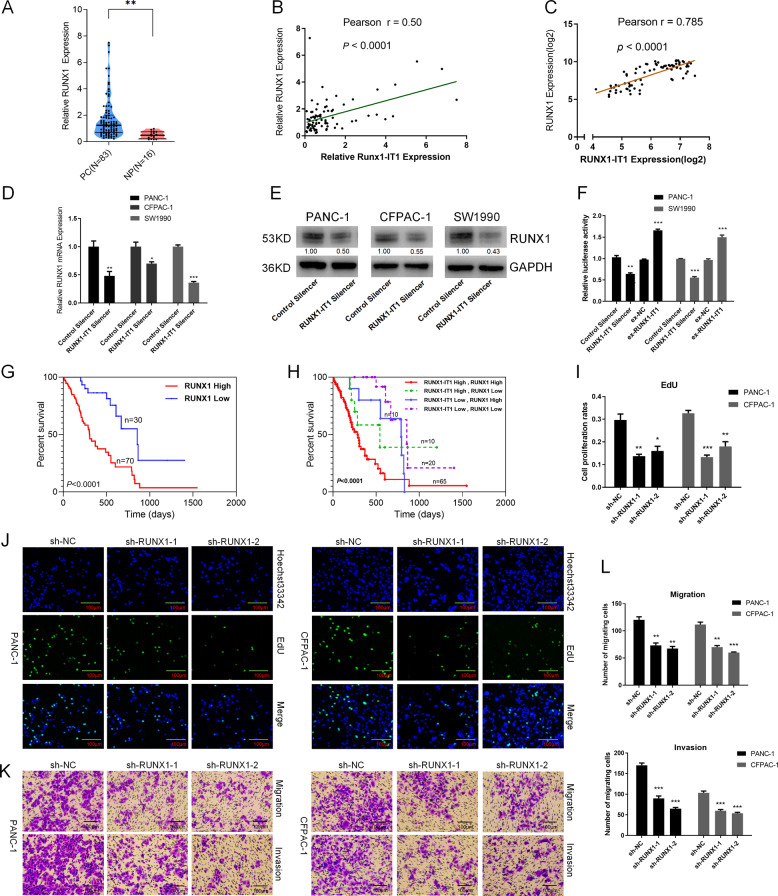
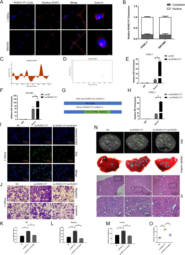
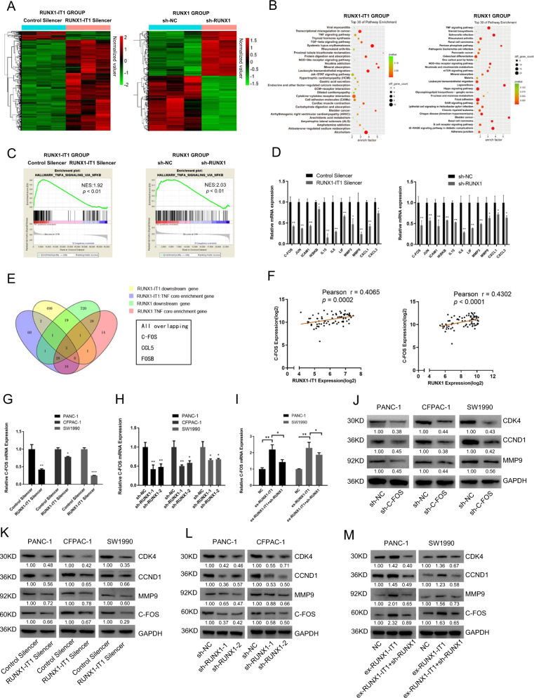
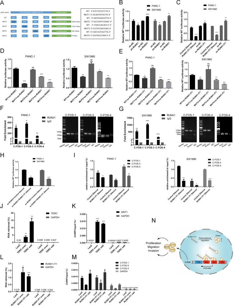
Numerous long noncoding RNAs (lncRNAs) are aberrantly expressed in pancreatic cancer (PC); however, their functions and mechanisms in cancer progression are largely unknown. In this study, we identified a novel PC-associated lncRNA, RUNX1-IT1, that was significantly upregulated in PC patient samples from multiple centers and associated with poor prognosis. In vitro and in vivo, alterations in RUNX1-IT1 expression markedly affected PC proliferation, migration and invasion. RUNX1-IT1 contributed to the progression of PC by interacting with the adjacent gene RUNX1. Rescue experiments showed that RUNX1 reduced the cancer-promoting effect of RUNX1-IT1. RNA-seq analysis after silencing RUNX1-IT1 and RUNX1 highlighted alterations in the common target C-FOS. Mechanistically, we demonstrated that RUNX1-IT1 was a trans-acting factor that participated in the proliferation, migration and invasion of PC by recruiting RUNX1 to the C-FOS gene promoter. Furthermore, RUNX1-IT1 enhanced the transcription of the RUNX1 gene, indicating its potential as a cis-regulatory RNA involved in the upstream regulation of RUNX1. Overall, RUNX1-IT1 is a crucial oncogenic lncRNA that activates C-FOS expression by regulating and recruiting RUNX1 and is a potential prognostic biomarker and therapeutic target for PC.


Conflict of interest statement

The authors declare that they have no conflict of interest.

Figures

Fig. 1
Fig. 1. LncRNA RUNX1-IT1 is overexpressed in PC.
a Hierarchical clustering analysis of genes that are differentially expressed in pancreatic cancer (PC) and normal pancreatic (NP) tissues (fold change: top 2000; P < 0.05). b Venn diagram of upregulated and downregulated genes in the three GEO datasets. c Expression profile of RUNX1-IT1 in the three GEO datasets. d RUNX1-IT1 expression was assessed by qRT-PCR in PC and NP tissues. e RUNX1-IT1 expression was assessed by qRT-PCR in paired PC tissues. f The RUNX1-IT1 expression in microarrays comprising PC (N = 175) and NP (N = 13) samples from three independent cohorts was assessed by ISH. g Kaplan–Meier analysis of the overall survival of PC patients based on the RUNX1-IT1 ISH score data. h Representative images of RUNX1-IT1-positive cells in PC and NP tissue samples from the three independent cohorts (scale bars, 500 and 50 μm) (*P < 0.05, **P < 0.01, ***P < 0.001).
Fig. 2
Fig. 2. Knockdown of RUNX1-IT1 significantly inhibits PC cell proliferation, migration and metastasis in vitro and in vivo.
a qRT-PCR analysis showed that RUNX1-IT1 expression was knocked down in PANC-1, CFPAC-1 and SW1990 cells by Smart Silencer lncRNA (RUNX1 Silencer and Control Silencer). b CCK8 assays were used to assess the viability of two groups of PC cells transfected with either RUNX1-IT1 Silencer or Control Silencer. c, d EdU assays were used to assess the cell proliferation ability. e, f Migration ability was assessed by a wound healing assay. g, h Transwell assays were used to assess the cell migration and invasion abilities. i Abdominal MRI scans of nude mice after 6 weeks of orthotopic PC xenograft growth. The livers of nude mice in the RUNX1-IT1 knockout PANC-1 cell group showed few metastatic lesions, but obvious metastatic lesions were found in the livers of the control group mice (CRISPR-Cas9 lentiviral system). j Images of micrometastases on the liver surface in the RUNX1-IT1 knockout PANC-1 group and in the control group. k HE staining of liver metastatic lesions was performed. The representative HE images are shown (scale bars, 1000 and 100 μm). l Histogram indicating the numbers of metastasized lesions in the two groups of mice. The number of metastatic lesions in the RUNX1-IT1 knockout PANC-1 group (N = 5) was significantly decreased compared with that in the control group (N = 5) (*P < 0.05, **P < 0.01, ***P < 0.001).
Fig. 3
Fig. 3. LncRNA RUNX1-IT1 expression is associated with the transcription factor RUNX1.
a RUNX1 expression was assessed by qRT-PCR in PC and NP tissues. b Correlation analysis of RUNX1-IT1 and RUNX1 expression in PC tissues by qPCR. c Correlation analysis of RUNX1-IT1 and RUNX1 using GEO data (GSE15471). d RUNX1 mRNA expression levels in RUNX1-IT1 knockdown and control cells were analyzed by qPCR. e The RUNX1 protein expression levels in RUNX1-IT1 knockdown and control cells were measured by WB. f Luciferase activity assays were performed on RUNX1-IT1 knockdown and RUNX1-IT1-overexpressing PANC-1 and SW1990 cells cotransfected with the pGL3 reporter vector containing the WT RUNX1 promoter (RUNX1 promoter length: 2200 bp from the transcription start site (TSS)). g Kaplan–Meier survival analysis of PC patients based on RUNX1 IHC score data. h Kaplan–Meier survival analysis of the four groups of PC patients based on RUNX1-IT1 ISH and RUNX1 IHC datas. i, j EdU assays were used to assess the proliferation ability of PANC-1 and SW1990 cells transfected with sh-RUNX1-1, sh-RUNX1-2, and control lentiviral vectors. k, l Transwell assays were used to assess the migration and invasion abilities of PANC-1 and SW1990 cells transfected with sh-RUNX1-1, sh-RUNX1-2 and control lentiviral vectors (*P < 0.05, **P < 0.01, ***P < 0.001).
Fig. 4
Fig. 4. RUNX1-IT1 functions via the transcription factor RUNX1 in PC.
a FISH analysis showing the subcellular localization of RUNX1-IT1 in PANC-1 and SW1990 cells. b Histogram showing the expression level of RUNX1-IT1 in the subcellular fractions of PANC-1 and SW1990 cells, as analyzed by qPCR. c The interaction profile, which represents the protein interaction score (Y axis) relative to the RUNX1-IT1 RNA sequence (X axis), provides information about the region most likely to be bound by the protein. d The interaction matrix, which shows a heatmap of the RUNX1 protein (Y axis) and RUNX1-IT1 RNA (X axis) regions. The red shading in the heatmap indicates the interaction score of a single amino acid and nucleotide pair. e, f RIP assays were performed to validate RUNX1-IT1 binding to RUNX1 in PANC-1 and SW1990 cells transfected with RUNX1-IT1 overexpression or control vectors. g The chart shows information about the WT and mutant RUNX1-IT1 vectors. h RIP assays were performed to validate RUNX1-IT1 binding to RUNX1 in PANC-1 cells transfected with RUNX1-IT1 (WT) and mutant RUNX1-IT1 overexpression vectors. i EdU assays were used to assess proliferation in the three groups of PANC-1 cells. j The migration and invasion abilities of the three groups of cells were assessed by a transwell assay. k Histogram showing the proliferation rates of cotransfected cells in the three groups. l, m Histogram showing the number of migrated and invaded cotransfected cells in the three groups. n Representative MRI images of liver metastatic tumors in the three groups of mice are shown. Images of liver surfaces and HE staining of metastatic tumor lesions in the different groups are shown (scale bars, 1000 and 100 μm). (o) Histogram indicating the numbers of metastasized lesions in the three groups of mice (*P < 0.05, **P < 0.01, ***P < 0.001).
Fig. 5
Fig. 5. C-FOS is a critical downstream target of RUNX1-IT1 and RUNX1.
a Heatmap of differentially expressed downstream genes in RUNX1 and RUNX1-IT1 knockdown PANC-1 cells and the corresponding control cells (P < 0.05, fold change > 2). b Top 30 enriched pathways in RUNX1 and RUNX1-IT1 knockdown PANC-1 cells. c GSEA analysis of the RUNX1 and RUNX1-IT1 knockdown PANC-1 groups and the control. d The levels of downstream TNF genes in the RUNX1 and RUNX1-IT1 knockdown PANC-1 groups were analyzed by qRT-PCR and compared with those in the control group. e The Venn diagram shows three targets in the area common to the four groups, namely, RUNX1-IT1 downstream genes, RUNX1 downstream genes (fold change > 2, P < 0.05), RUNX1-IT1 TNF core enriched genes and RUNX1 TNF core enriched genes (P < 0.05). f Correlation analysis between C-FOS and RUNX1 and between C-FOS and RUNX1-IT1 in GEO (GSE15471). g, h The C-FOS gene expression levels in the RUNX1-IT1 knockdown and RUNX1 knockdown groups were analyzed by qPCR. i The C-FOS gene expression levels in the NC, ex-RUNX1-IT1 and ex-RUNX1-IT1+sh-RUNX1 groups were analyzed by qRT-PCR. j The protein expression levels of C-FOS-related downstream molecules in the C-FOS knockdown PC and control cells were assessed by WB. k, l The protein expression levels of C-FOS and its related downstream molecules in the RUNX1-IT1 knockdown and RUNX1 knockdown groups were assessed by WB. m The protein expression levels of C-FOS and its related downstream molecules in the NC, ex-RUNX1-IT1 and ex-RUNX1-IT1 + sh-RUNX1 groups were assessed by WB (*P < 0.05, **P < 0.01, ***P < 0.001).
Fig. 6
Fig. 6. RUNX1-IT1 and RUNX1 promoted cell proliferation, migration and invasion via C-FOS.
a, c Knockdown of C-FOS in RUNX-IT1-overexpressing cells. EdU and transwell assays were used to assess proliferation, migration and invasion in the NC, ex-RUNX1-IT1 and ex-RUNX1-IT1+sh-C-FOS groups of PANC-1 cells. b, d Knockdown of C-FOS in RUNX1-overexpressing cells. EdU and transwell assays were used to assess proliferation, migration and invasion in the NC, ex-RUNX1 and ex-RUNX1+sh-C-FOS groups of PANC-1 cells. e Histogram showing the proliferation rates of cotransfected cells. f, g Histogram showing the numbers of migrated and invaded cotransfected cells. h The protein expression levels of C-FOS-related downstream molecules in the NC, ex-RUNX1-IT1 and ex-RUNX1-IT1+sh-C-FOS groups were assessed by WB. i The protein expression levels of C-FOS-related downstream molecules in the NC, ex-RUNX1 and ex-RUNX1+sh-C-FOS groups were assessed by WB analysis (*P < 0.05, **P < 0.01, ***P < 0.001).
Fig. 7
Fig. 7. RUNX1-IT1 expression is positively associated with RUNX1 and C-FOS expression in human PC.
a Representative images (scale bars, 500 and 50 μm) of ISH or IHC staining for RUNX1-IT1, RUNX1, C-FOS, MMP9 and CCND1 in tissues from different clinical stages (I–IV) are shown. be Bar charts showing the correlation between the expression of RUNX1-IT1 and RUNX1, C-FOS, MMP9 and CCND1. f General correlation analysis of RUNX1-IT1, RUNX1, C-FOS, MMP9 and CCND1. (*P < 0.05, **P < 0.01, ***P < 0.001).
Fig. 8
Fig. 8. RUNX1-IT1 regulates C-FOS expression by recruiting RUNX1 to the C-FOS promoter.
a WT and mutant C-FOS promoters were constructed using the pGL3 vector. b, c Luciferase activity assays were performed using PC cells (PANC-1 and SW1990) with RUNX1 or RUNX1-IT1 knockdown and overexpression that were cotransfected with a C-FOS WT promoter. d, e RUNX1- or RUNX1-IT1-overexpressing PC cells were cotransfected with the WT vector or with one of the MUT vectors. f, g ChIP assays with an anti-RUNX1 antibody or IgG were performed to verify the enrichment of RUNX1 at binding sites in the C-FOS promoter in PC cells. The ChIP-PCR products of the RUNX1, input and IgG groups were detected by agarose gel electrophoresis. h RUNX1-overexpressing PC cells were transfected with RUNX1-IT1 or Control Silencer. Luciferase activity assays were performed. i ChIP assays were performed in RUNX1-IT1 knockdown PC and control cells with an anti-RUNX1 antibody or IgG. j Retrieval of RNA by ChIRP with TERC probes (positive control). ChIRP was performed using PANC-1 cells and TERC lncRNA even, odd or NC (LacZ) probe sets. Purified RNA was then analyzed by qRT-PCR using RNA-positive control primers (TERC) and RNA-negative control primers (GAPDH). k Successful DNA binding by ChIRP with TERC probes. Purified DNA was then analyzed by qRT-PCR using the WNT-1 precursor (positive target) and GAPDH D2 coding region (negative target). l Successful retrieval of RNA by ChIRP with RUNX1-IT1 probes. m DNA binding by ChIRP with the RUNX1-IT1 probe. Purified DNA was analyzed by qRT-PCR using primers specific for the FOS promoter binding region and the GAPDH D2 coding region. n A schematic model of the mechanism underlying the role of RUNX1-IT1 in the progression of PC (ns no significance, *P < 0.05, **P < 0.01, ***P < 0.001).

Similar articles

Cited by

References

    1. Siegel RL, Miller KD, Jemal A. Cancer statistics, 2018. CA Cancer J. Clin. 2018;68:7–30. doi: 10.3322/caac.21442. - DOI - PubMed
    1. Rahib L, et al. Projecting cancer incidence and deaths to 2030: the unexpected burden of thyroid, liver, and pancreas cancers in the United States. Cancer Res. 2014;74:2913–2921. doi: 10.1158/0008-5472.CAN-14-0155. - DOI - PubMed
    1. Habib J, Yu J. Circulating tumor cells in pancreatic cancer. J. Pancreatol. 2019;2:54–59. doi: 10.1097/JP9.0000000000000014. - DOI
    1. Puleo F, et al. New challenges in perioperative management of pancreatic cancer. World J. Gastroenterol. 2015;21:2281–2293. doi: 10.3748/wjg.v21.i8.2281. - DOI - PMC - PubMed
    1. Zhan HX, Xu JW, Wu D, Zhang TP, Hu SY. Pancreatic cancer stem cells: new insight into a stubborn disease. Cancer Lett. 2015;357:429–437. doi: 10.1016/j.canlet.2014.12.004. - DOI - PubMed

Publication types

MeSH terms

Substances