. 2020 Feb;30(2):133-145.
doi: 10.1038/s41422-019-0264-2. Epub 2019 Dec 18.

Dosage effect of multiple genes accounts for multisystem disorder of myotonic dystrophy type 1

Affiliations

Dosage effect of multiple genes accounts for multisystem disorder of myotonic dystrophy type 1

Qi Yin et al. Cell Res. 2020 Feb.

Abstract

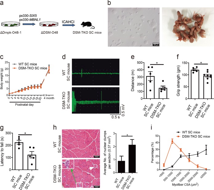
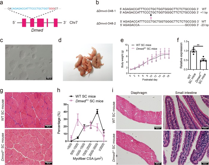
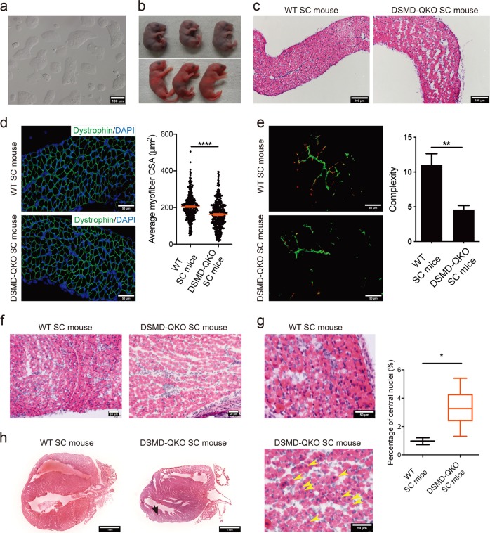
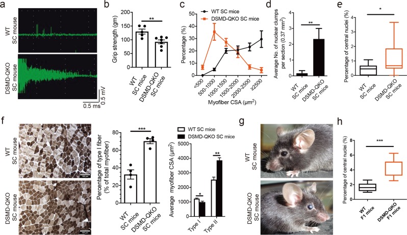
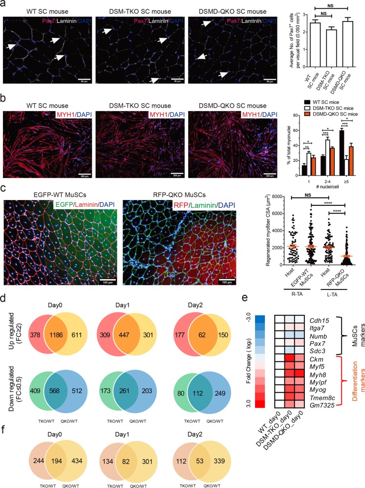
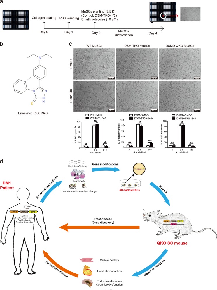
Multisystem manifestations in myotonic dystrophy type 1 (DM1) may be due to dosage reduction in multiple genes induced by aberrant expansion of CTG repeats in DMPK, including DMPK, its neighboring genes (SIX5 or DMWD) and downstream MBNL1. However, direct evidence is lacking. Here, we develop a new strategy to generate mice carrying multigene heterozygous mutations to mimic dosage reduction in one step by injection of haploid embryonic stem cells with mutant Dmpk, Six5 and Mbnl1 into oocytes. The triple heterozygous mutant mice exhibit adult-onset DM1 phenotypes. With the additional mutation in Dmwd, the quadruple heterozygous mutant mice recapitulate many major manifestations in congenital DM1. Moreover, muscle stem cells in both models display reduced stemness, providing a unique model for screening small molecules for treatment of DM1. Our results suggest that the complex symptoms of DM1 result from the reduced dosage of multiple genes.


Conflict of interest statement

The authors declare no competing interests.

Figures

Fig. 1
Fig. 1
SC mice carrying heterozygous mutations in Dmpk, Six5 and Mbnl1 exhibit typical DM1-associated muscle defects. a Diagram of DSM-TKO SC mice generated by ICAHCI of ΔDSM-O48 cells carrying mutatnt Dmpk, Six5 and Mbnl1. b Images of cultured ΔDSM-O48-1 haploid ESCs and newborn SC pups from ΔDSM-O48-1 cells. Scale bar, 100 μm. c Body weight analysis of DSM-TKO and WT SC mice (n > 5 per group, means ± SD). d EMG analysis of TKO SC mice (WT SC mice, n = 3; DSM-TKO SC mice, n = 2, 4-month old). e-g Muscle weakness of DSM-TKO SC mice was determined by treadmill test (e) (n = 5 per group, 4-month old), forelimb grip strength test (f) (WT SC mice, n = 5; DSM-TKO SC mice, n = 8, 4-month old) and rotarod test (g) (n = 5 per group, 4-month old). Unpaired Student’s t-test, *P < 0.05. h H&E staining of TA muscles from DSM-TKO and WT SC mice, DSM-TKO mice showing nuclear clump (green arrow) and atrophic fiber (white arrow) (WT SC mice, n = 11; DSM-TKO SC mice, n = 7, 4-month old). Unpaired Student’s t-test, *P < 0.05. Scale bars, 50 μm. i TA myofiber CSA analysis (n = 3 per group, 4-month old)
Fig. 2
Fig. 2
Generation of Dmwd+/ SC mice through ICAHCI of haploid cells carrying mutant Dmwd. a Schematic of the sgRNA targeting Dmwd. b Sequences of the Dmwd gene in two cell lines (ΔDmwd-O48-1 and ΔDmwd-O48-2) carrying CRISPR-Cas9-induced gene modifications. c Phase-contrast image of ΔDmwd-O48-1 cell line. Scale bar, 100 μm. d Newborn SC pups generated from ΔDmwd-O48-1 cells. e Average body weight of Dmwd+/ SC mice and WT SC mice (n > 8 per group, means ± SD). f Transcription analysis of Dmwd in TA muscles showed that the transcription level of Dmwd was significantly lower in Dmwd+/ SC mice compared with WT SC mice (n = 4 per group, 4-6-month old). Unpaired Student’s t-test, **P < 0.01. g Representative images of H&E staining of TA muscles from DMWD+/ SC mice and WT SC mice (4-6-month old). Scale bars, 50 μm. h CSA analysis of TA muscle showed muscle wasting in DMWD+/ SC mice (n = 3 per group, 4-6-month old). i Representative images of H&E staining showed normal histological structure of diaphragm and small intestine in DMWD+/ SC mice (4-6-month old). Scale bars, 200 µm for diaphragm; 50 µm for small intestine
Fig. 3
Fig. 3
SC mice carrying heterozygous mutations in Dmpk, Six5, Mbnl1 and Dmwd show CDM phenotypes. a Phase-contrast image of cultured ΔDSMD-O48-1 haploid cells. Scale bar, 100 μm. b 23% of newborn DSMD-QKO SC pups died during perinatal period (upper), while others survived (lower). c H&E staining of diaphragm sections from DSMD-QKO and WT SC pups (P1). Scale bars, 100 μm. d Diaphragm CSA analysis of DSMD-QKO and WT SC mice (P1) (n = 3 per group). Unpaired Student’s t-test, ****P< 0.0001. Scale bars, 50 μm. e Immunofluorescent staining of bungarotoxin (red) and neurofilament H (green), and complexity analysis of NMJs of DSMD-QKO and WT SC mice (P1) (WT SC pups, n = 2; DSMD-QKO SC pups, n = 5). Unpaired Student’s t-test, **P< 0.01. Scale bars, 50 μm. f H&E staining of TA muscle sections (P1) showing less myofiber in DSMD-QKO SC mice (2/5). Scale bars, 50 μm. g H&E staining of TA muscle of DSMD-QKO and WT SC (P0) mice and the percentage of myofibers containing centrally located nuclei in TA muscle (n = 3 per group). Yellow arrows indicate the myofibers with central nuclei. Unpaired Student’s t-test, *P < 0.05. Scale bars, 50 μm. h H&E staining of heart cryo-sections (P17) showing left ventricular posterior wall attenuation in DSMD-QKO SC mice (2/7, black arrow). Scale bars, 1 mm
Fig. 4
Fig. 4
DM1-related pathogenic phenotypes in adult SC mice carrying quadruple mutations. a EMG analysis indicates typical waxing-waning myotonia in QKO SC mice (WT SC mice, n = 3; DSMD-QKO SC mice, n = 5, 4-6-month old). b Forelimb grip strength test (WT SC mice, n = 5; DSMD-QKO SC mice, n = 7, 4-6-month old). Unpaired Student’s t-test, **P< 0.01. c TA CSA analysis (n = 3 per group, 4-6-month old). d Nuclear clump analysis of TA muscle (WT SC mice, n = 6; DSMD-QKO SC mice, n = 3, 4-6-month old). Unpaired Student’s t-test, **P< 0.01. e The percentage of myofibers containing centrally located nuclei in TA muscle of QKO and WT SC mice (P30) (n = 3 per group). Unpaired Student’s t-test, *P < 0.05. f Representative images of ATPase staining and fiber type analysis (n = 4 per group, 4-6-month old). Unpaired Student’s t-test, *P< 0.05, **P< 0.01, ***P< 0.001. Scale bars, 100 μm. g Dust-like opacities in eyes of DSMD-QKO SC mice (3/7, 4-6-month old). h The percentage of myofibers containing centrally located nuclei in TA muscles of QKO and WT F1 mice (n = 5 per group, P0). Unpaired Student’s t-test, ***P < 0.001
Fig. 5
Fig. 5
Decreased differentiation potential of MuSCs from TKO or QKO DM1 mice. a Representative images of PAX7 immunostaining (pink) in TA muscle sections from adult TKO and QKO mice (4-6-month old, n = 3 per group). White arrows indicate PAX7-positive cells. Scale bars, 50 µm. Unpaired Student’s t-test. NS, no significant differences. b In vitro differentiation of MuSCs (n = 3 per group, 2-month old). One-way ANOVA, *P< 0.05, **P< 0.01, ***P< 0.001. Scale bars, 200 μm. c In vivo differentiation of EGFP-WT MuSCs and RFP-QKO MuSCs (n = 2 per group, 2-month old). R-TA represents TA of right hind leg, while L-TA represents TA of left hind leg. One-way ANOVA, ****P< 0.0001. NS, no significant differences. Scale bar, 100 μm. d Venn diagrams show that the number of common DEGs between mutant and WT samples dramatically decreased following MuSC differentiation (from Day 0 to Day 2). e Heat map of stem cell markers and differentiation markers in DM1 and WT MuSCs. f Venn diagrams show that the number of common mis-splicing genes (skipped exon events) between mutant and WT samples dramatically decreased following MuSC differentiation (from Day 0 to Day 2)
Fig. 6
Fig. 6
Large-scale screening and potential applications of DM1 mice. a Diagram of large-scale screening for small molecules that can promote differentiation of DM1 MuSCs. b Molecular structure of T5381948. c Images show that T5381948 could specifically improve the in vitro differentiation of DSM-TKO MuSCs. Scale bars, 200 μm. Unpaired Student’s t-test; ***P< 0.001; NS, no significant differences. d Overview of the generation and applications of DM1 mouse model with quadruple heterozygous mutations

Comment in

Similar articles

Cited by

References

    1. Korade-Mirnics Z, Babitzke P, Hoffman E. Myotonic dystrophy: molecular windows on a complex etiology. Nucleic Acids Res. 1998;26:1363–1368. doi: 10.1093/nar/26.6.1363. - DOI - PMC - PubMed
    1. Ranum LP, Day JW. Pathogenic RNA repeats: an expanding role in genetic disease. Trends Genet. 2004;20:506–512. doi: 10.1016/j.tig.2004.08.004. - DOI - PubMed
    1. Udd B, Krahe R. The myotonic dystrophies: molecular, clinical, and therapeutic challenges. Lancet Neurol. 2012;11:891–905. doi: 10.1016/S1474-4422(12)70204-1. - DOI - PubMed
    1. Brook JD, et al. Molecular basis of myotonic dystrophy: expansion of a trinucleotide (CTG) repeat at the 3' end of a transcript encoding a protein kinase family member. Cell. 1992;68:799–808. doi: 10.1016/0092-8674(92)90154-5. - DOI - PubMed
    1. Mahadevan M, et al. Myotonic dystrophy mutation: an unstable CTG repeat in the 3' untranslated region of the gene. Science. 1992;255:1253–1255. doi: 10.1126/science.1546325. - DOI - PubMed

Publication types