. 2020 Mar;21(3):330-348.
doi: 10.1111/mpp.12899. Epub 2020 Jan 8.

Extensive chromosomal rearrangements and rapid evolution of novel effector superfamilies contribute to host adaptation and speciation in the basal ascomycetous fungi

Affiliations

Extensive chromosomal rearrangements and rapid evolution of novel effector superfamilies contribute to host adaptation and speciation in the basal ascomycetous fungi

Qinhu Wang et al. Mol Plant Pathol. 2020 Mar.

Abstract

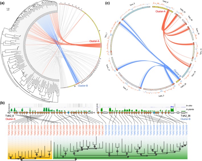
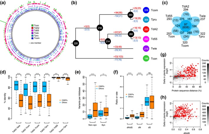
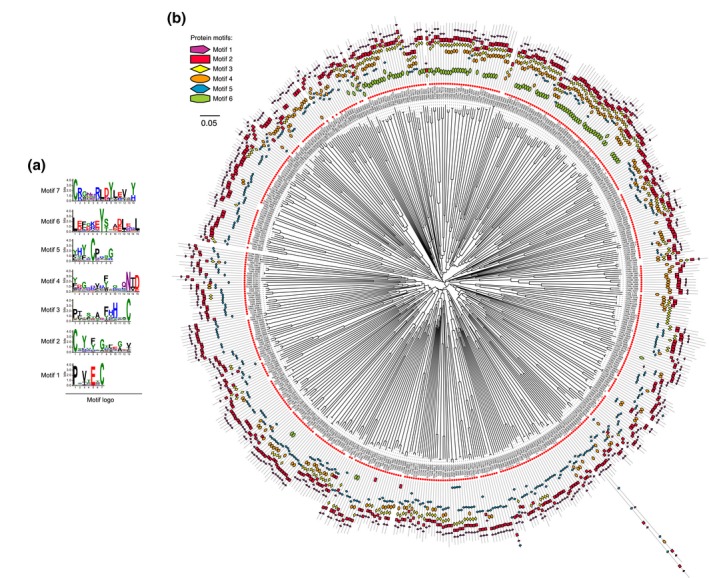
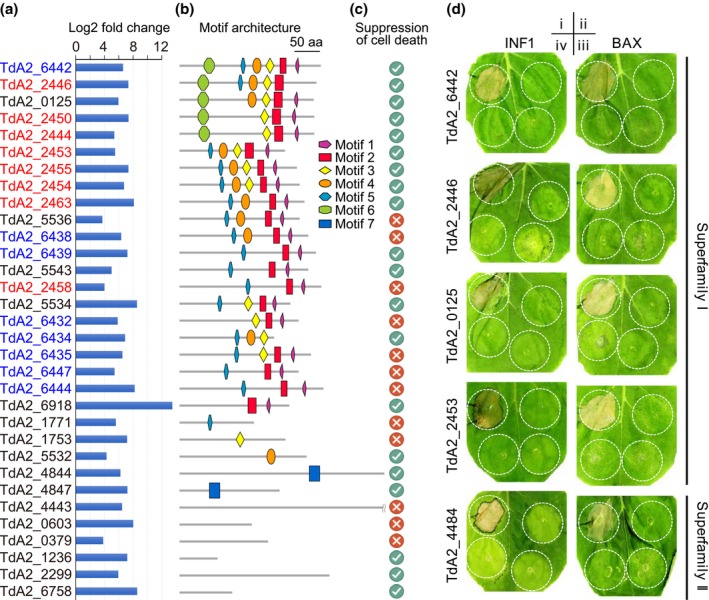
The basal ascomycetes in genus Taphrina have strict host specificity and coevolution with their host plants, making them appealing models for studying the genomic basis of ecological divergence and host adaption. We therefore performed genome sequencing and comparative genomics of different Taphrina species with distinct host ranges to reveal their evolution. We identified frequent chromosomal rearrangements and highly dynamic lineage-specific (LS) genomic regions in Taphrina genomes. The LS regions occur at the flanking regions of chromosomal breakpoints, and are greatly enriched for DNA repeats, non-core genes, and in planta up-regulated genes. Furthermore, we identified hundreds of candidate secreted effector proteins (CSEPs) that are commonly organized in gene clusters that form distinct AT-rich isochore-like regions. Nearly half of the CSEPs constitute two novel superfamilies with modular structures unique to Taphrina. These CSEPs are commonly up-regulated during infection, enriched in the LS regions, evolved faster, and underwent extensive gene gain and loss in different species. In addition to displaying signatures of positive selection, functional characterization of selected CSEP genes confirmed their roles in suppression of plant defence responses. Overall, our results showed that extensive chromosomal rearrangements and rapidly evolving CSEP superfamilies play important roles in speciation and host adaptation in the early-branching ascomycetous fungi.

Keywords: Taphrina; adaptation; comparative genomics; fungi; host specificity; secreted effector protein; speciation.


Figures

Figure 1
Figure 1
Biological features of Taphrina deformans on peach (Prunus persica) leaves. (a) Peach leaf curl symptom caused by T. deformans. (b) Yeast cells of histone H1‐GFP transformant of T. deformans A2 (TdA2) were examined by light and epifluorescence microscopy. (c) Asci with ascospore formed on the surface of diseased peach leaves. (d, e) Light and electron microscopy of biotrophic hyphae (marked with arrows) of TdA2 growing in the extracellular spaces of peach leaf cells. (f) Dikaryotic hyphae of histone H1‐GFP transformant of TdA2 grown in peach leaves were examined by light and epifluorescence microscopy. Bars, 10 μm
Figure 2
Figure 2
Phylogenomic relationship, genome features, and host specificity of sequenced Taphrina species. (a) Neighbour‐joining phylogenomic tree constructed with a concatenated set of 4,802 single‐copy orthologue families conserved in the sequenced Taphrina genomes (*, sequenced in this study). All nodes have a 100% bootstrap support. Scale bar corresponds to 0.05 amino acid substitutions per site. The number of gained (+) and lost (−) orthologue families was estimated on each branch of the tree under a birth–death evolutionary model. Figures in circles are the exact number of orthologue families in the nodes. TdA2, T. deformans strain A2; Td55, T. deformans strain CBS 355.35; TdJCM, T. deformans strain JCM 22205; Twie, T. wiesneri strain CBS 275.28; TwJCM, T. wiesneri strain JCM 22204; Tcom, T. communis strain CBS 352.35; Tpru, T. pruni strain CBS 358.35; Tcon, T. confusa strain CBS 375.39; Tfla, T. flavorubra strain JCM 22207; Tpop, T. populina strain CBS 337.55. (b) Genome size and number of predicted protein‐coding genes. (c) Percentage of four marked categories of repeats. (d) Number of protein‐coding genes belonging to orthologue families conserved in all (Core) or a subset (Shared) of Taphrina species or species/strain‐specific (Unique). (e) Number of predicted carbohydrate‐active enzymes (CAZY), secreted proteins (Secretome), and candidate secreted effector proteins (CSEPs). (f) Host species and phylogeny. The tree was manually drawn based on previous studies (Lee and Wen, 2001; Mowrey and Werner, 1990; Wen et al., 2008). Amy, Ce, Pad, and Pr stand for subgenera Amygdalus (almonds and peaches), Cerasus (cherries), Padus (bird cherries), and Prunus (plums and apricots), respectively
Figure 3
Figure 3
Chromosomal evolution of Taphrina strains. (a) Examples of chromosomal rearrangement events in Taphrina. Syntenic chromosomal regions are marked with the same colour and connected with sectors. +, Watson strand; −, Crick strand. Red dots indicate telomeric repeat. (b) Chromosomes of marked Taphrina strains were separated by pulsed‐field gel electrophoresis. Chromosomes of Saccharomyces cerevisiae YPH80 were the molecular weight markers (M). Each strain has more than 17 chromosomal bands, ranging from 225 to 815 kb. *Bands probably containing more than one chromosome. (c) Violin‐plot showing the enrichment of DNA repeat in the lineage‐specific (LS) genomic regions of Taphrina. (d) Violin‐plot showing the depletion and enrichment of core and noncore genes, respectively, in the LS genomic regions of Taphrina. (e) Bar‐plot showing the enrichment of genes up‐regulated during infection in the LS genomic regions of TdA2. *p < .05; **p < .01; ns, not significant. The statistical significances were accessed by one‐sided Wilcoxon tests (c, d) and Fisher's exact tests (e)
Figure 4
Figure 4
Intraspecies genomic variations among Taphrina deformans strains. (a) Circos plot of genome features of T. deformans. Concentric circles show different features along the 37 largest scaffolds of TdA2 that were drawn in 10 kb non‐overlapping windows: a, ideograms of the top 37 scaffolds; b, heatmap of gene density; c, heatmap of repeat density; d, distribution of candidate secreted effector protein (CSEP) genes; e, RNA‐Seq read coverages of the in planta samples (pathogenic phase, pink) and yeast cells (saprophytic phase, green) on a log2 scale; f–h, distribution of variants from TdJCM, Td55, and Td56, respectively, in comparison with TdA2; i, GC content plotted as the deviation (higher, green; lower, red) from the average GC content of the entire genome; j, genes derived from intragenomic duplications are connected by green lines. (b) Neighbour‐joining phylogenetic tree of the four T. deformans strains constructed with MEGA 6 (Tamura et al., 2013) based on genome‐wide single nucleotide variant sites. Scale bar corresponds to one nucleotide substitution per site. (c) Venn diagram of shared and unique variant sites among different T. deformans strains. The number outside the Venn diagram shows the total number of variants for each strain compared to TdA2. (d) Histogram of variant density distributions for T. deformans based on a bin of 10 kb. The curves illustrate the distributions estimated based on a two‐component mixture model using the expectation‐maximization algorithm. The mean and standard deviation values for the two curves are indicated. (e) Boxplot comparing the ratio of the number of nonsynonymous sites (non‐syn) to the number of synonymous sites (syn) per gene in the fast‐ and slow‐evolving genomic regions. ****p < .0001, t test. (f) Percentage of genes with over 2‐fold changes in expression in planta in the fast‐ and slow‐evolving genomic regions. ****p < .0001, χ2 test. (g) Distributions of gene border lengths in strain TdA2. The gene density is measured by gene borders, that is, the 5′ and 3′ flanking intergenic regions (FIR) lengths of the gene. The x axis and y axis are the logarithm of 5′ FIR and 3′ FIR, respectively. (h) The average number of repeats in the fast and slow‐evolving genomic regions. ns, not significant, χ2 test
Figure 5
Figure 5
Evolution of candidate secreted effector protein (CSEP) gene clusters in Taphrina. (a) Correlation between phylogeny and genomic location of CSEP genes in Taphrina deformans strain A2 (TdA2). Semicircular neighbour‐joining phylogenetic tree displays the relationships of CSEPs. Ideograms of the scaffolds containing CSEP genes are proportional to their sizes except the scaffolds 8 and 36, which contain the two largest CSEP gene clusters (Cluster A and Cluster B) and are enlarged and highlighted in yellow. The locations of all CSEP genes are indicated by red bars. Each gene of the tree is linked to its position on chromosomes. The lines starting from the highlighted scaffolds 8 and 36 are coloured in red and blue, respectively. (b) Phylogenetic relationship and genomic location of CSEP genes in the two largest clusters of TdA2. The p values of SH‐aLRT are plotted as circles on the branches with the circle size proportional to the p value (p > .5 only). Correlations between the phylogenetic relationships of CSEP genes and their locations on the genomic sequences (scaffolds 8 and 36) are connected by lines. Genes encoding CSEPs, CSEP‐orthologues without a detectable signal peptide, and nonsecreted proteins are shaded in orange, blue, and grey, respectively. Normalized RNA‐Seq read coverages derived from the in planta samples (green) and yeast cells in vitro (blue) are shown above. (c) Circos plot showing the conservation of the two largest CSEP gene clusters between different Taphrina genomes. The location of each CSEP gene is indicated by a red bar. Putatively orthologous CSEP genes are connected with lines. The lines starting from scaffolds 8 and 36 of TdA2 are coloured red and blue, respectively. The deviation from the average GC content of the entire scaffold is shown in the outside of the ideograms
Figure 6
Figure 6
Evolution of candidate secreted effector protein (CSEP) genes in Taphrina. (a–c) Gain and loss of the CSEP genes in different Taphrina species. (a) Expansion and contraction of the members of CSEP orthologue families in different Taphrina species. The multibars in the ring of the circular plot showing the number of CSEP members in each orthologue family. For a given orthologue family, the height of a bar is proportional to the number of CSEP members in the species, while the absence of a bar means that the CSEP orthologue family was lost in the species. (b) Evolution of Taphrina CSEP gene repertoire. The number of gained (in red) and lost (in blue) CSEP orthologue families (or CSEP genes in parentheses) was estimated on each branch of the tree under a birth–death evolutionary model. Figure in circles are the exact number of CSEP orthologue families in the nodes. (c) Venn diagram of shared and unique CSEP orthologue families. Figures in parentheses indicate numbers of genes in orthologue families. Figures outside the Venn diagram show the total number of CSEP orthologues in each genome. (d–h) Elevated evolutionary rate and induced expression of CSEPs in Taphrina pathogens. (d) Sequence identity between orthologous pairs of CSEP and other (non‐CSEP) proteins in marked Taphrina species. (e) Density of synonymous and nonsynonymous nucleotide variants between orthologous genes encoding CSEPs and other proteins in T. deformans. (f) The nonsynonymous (dN) or synonymous (dS) substitution rate and dN/dS ratio of orthologous genes encoding CSEPs and other proteins in the same Taphrina species as (a). (g) Two‐dimensional histogram of the protein sequence distance (1, identity %) between orthologous pairs and log2 fold change of in planta gene expression. CSEP genes are marked as red dots. (h) Two‐dimensional histogram of the dN/dS ratio and log2 fold change of in planta gene expression. CSEP genes are marked as red dots
Figure 7
Figure 7
Conserved motifs and the motif architecture of the superfamily I candidate secreted effector proteins (CSEPs). (a) Sequence logos show the six conserved motifs of superfamily I CSEPs that were identified de novo by MEME. (b) Modular structure and relationship of Taphrina superfamily I CSEPs. The circular neighbour‐joining dendrogram displays the modular structure and relationships of the members of CSEP orthologue families from marked Taphrina species with motifs 1 to 6 identified in this study. The motif architecture for each sequence is depicted in the outer ring. Red circles indicate a protein harbouring a signal peptide (CSEPs)
Figure 8
Figure 8
Candidate secreted effector proteins (CSEPs) suppress plant defence response. (a) The up‐regulation expression level of the selected representative CSEP genes used for functional analysis. The CSEPs in red and blue are from Cluster A and Cluster B, respectively. (b) The motif architecture of the selected CSEPs. (c) The ability of the selected CSEPs to suppress plant cell death. The ticks and crosses indicate that CSEP could or could not suppress both INF1‐ and BAX‐triggered plant cell deaths, respectively. (d) Examples showing the suppressions of INF1‐ or BAX‐triggered cell death in Nicotiana benthamiana by marked Taphrina CSEPs. The rest of the cell death assays are shown in Figure S9. N. benthamiana leaves were injected with Agrobacterium tumefaciens GV3101 expressing INF1 (left column) or BAX (right column) gene only (i), CSEP gene only (ii), or infiltration with agrobacterial cells expressing INF1 or BAX gene 12 hr (iii) or 16 hr (iv) after infiltration with cells expressing marked CSEP genes

Similar articles

Cited by

References

    1. Agrios, G.N. (2005) Plant Pathology, 5th edition Amsterdam, Netherlands: Academic Press.
    1. Ames, R.M. , Money, D. , Ghatge, V.P. , Whelan, S. and Lovell, S.C. (2012) Determining the evolutionary history of gene families. Bioinformatics, 28, 48–55. 10.1093/bioinformatics/btr592 - DOI - PubMed
    1. Bailey, T.L. , Boden, M. , Buske, F.A. , Frith, M. , Grant, C.E. , Clementi, L. et al (2009) MEME suite: tools for motif discovery and searching. Nucleic Acids Research, 37, W202–208. 10.1093/nar/gkp335 - DOI - PMC - PubMed
    1. Benaglia, T. , Chauveau, D. , Hunter, D.R. and Young, D. S. (2009) mixtools: an R package for analyzing finite mixture models. Journal of Statistical Software, 32, 1–29.
    1. Boetzer, M. , Henkel, C.V. , Jansen, H.J. , Butler, D. , and Pirovano, W. (2011) Scaffolding pre‐assembled contigs using SSPACE. Bioinformatics, 27, 578–579. 10.1093/bioinformatics/btq683 - DOI - PubMed

Publication types