. 2021 Jan 6;29(1):103-120.
doi: 10.1016/j.ymthe.2020.09.037. Epub 2020 Oct 1.

ESC-sEVs Rejuvenate Aging Hippocampal NSCs by Transferring SMADs to Regulate the MYT1-Egln3-Sirt1 Axis

Affiliations

ESC-sEVs Rejuvenate Aging Hippocampal NSCs by Transferring SMADs to Regulate the MYT1-Egln3-Sirt1 Axis

Guowen Hu et al. Mol Ther. .

Abstract

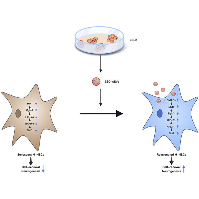
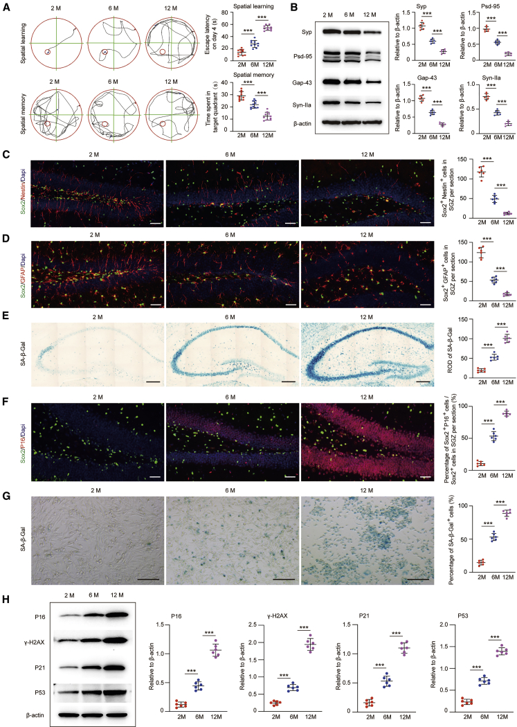
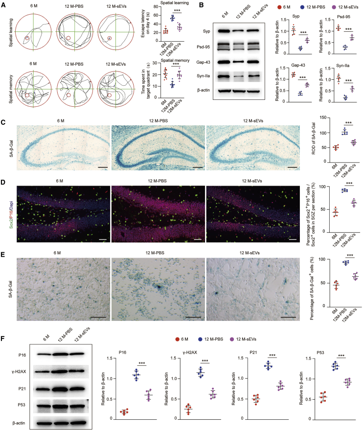
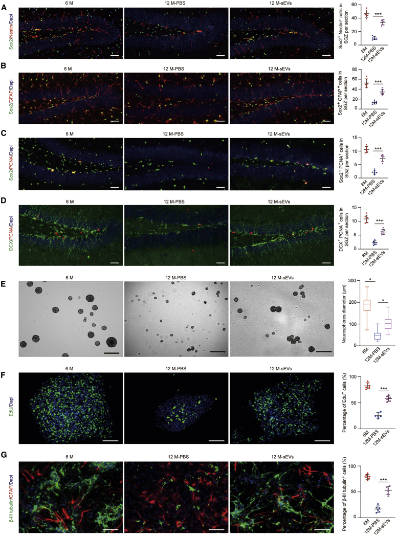
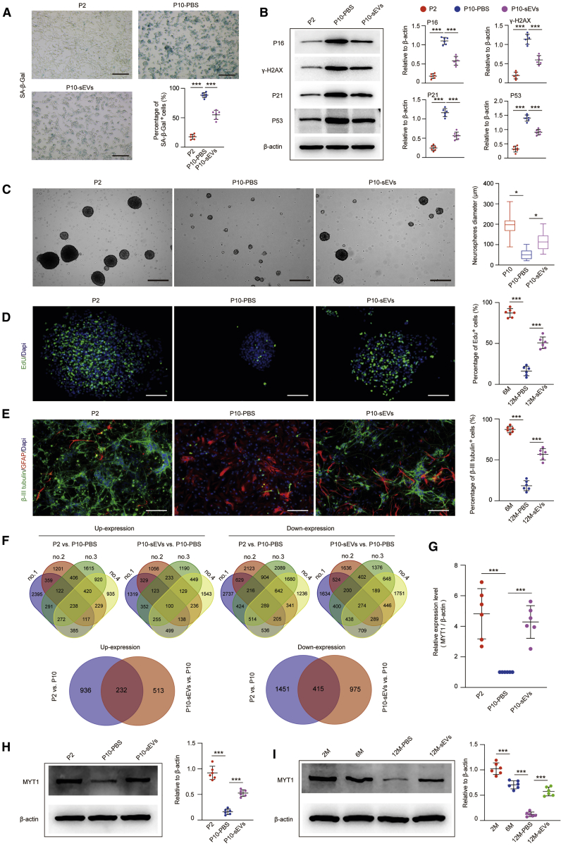
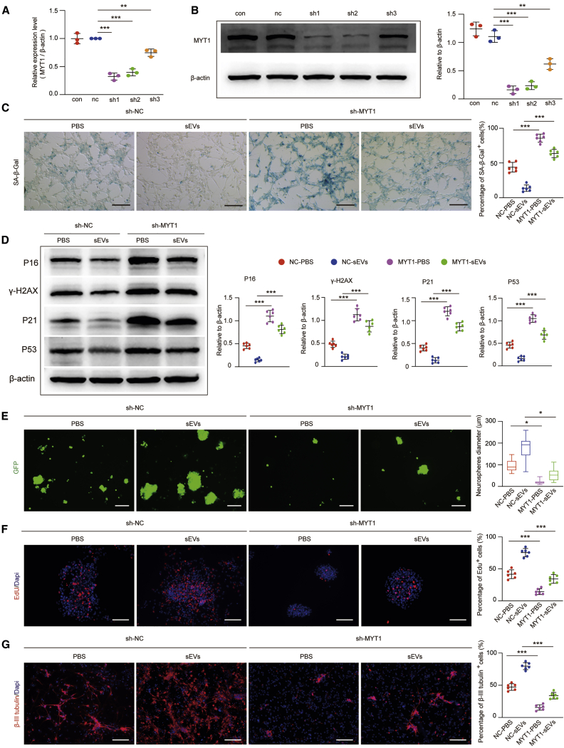
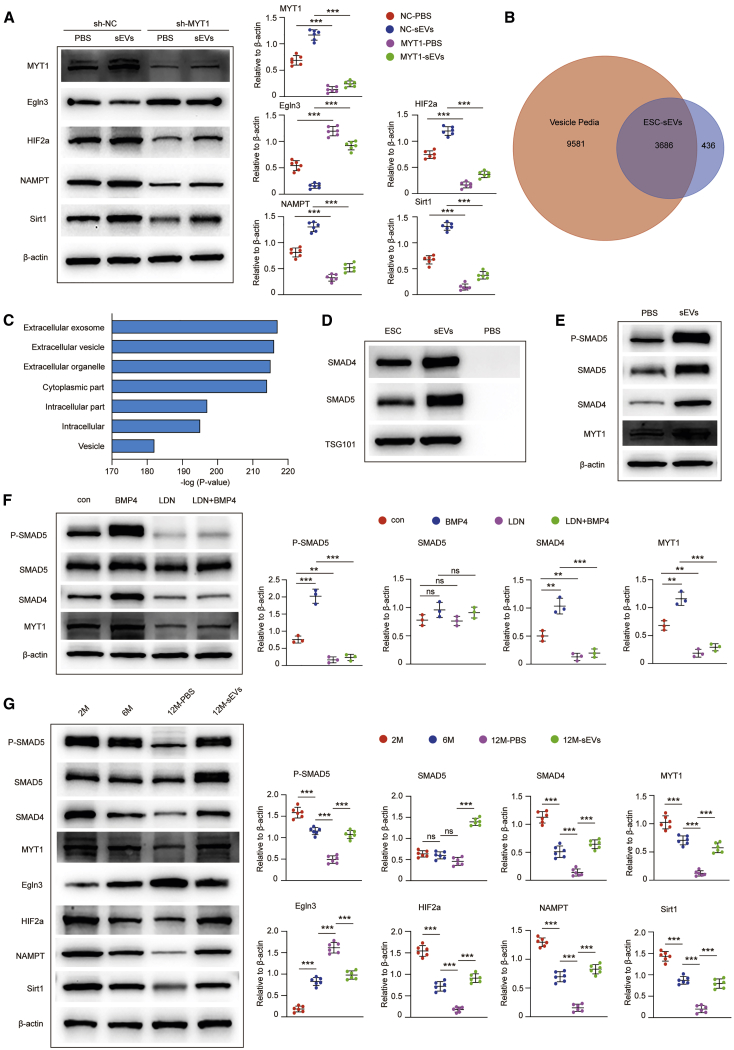
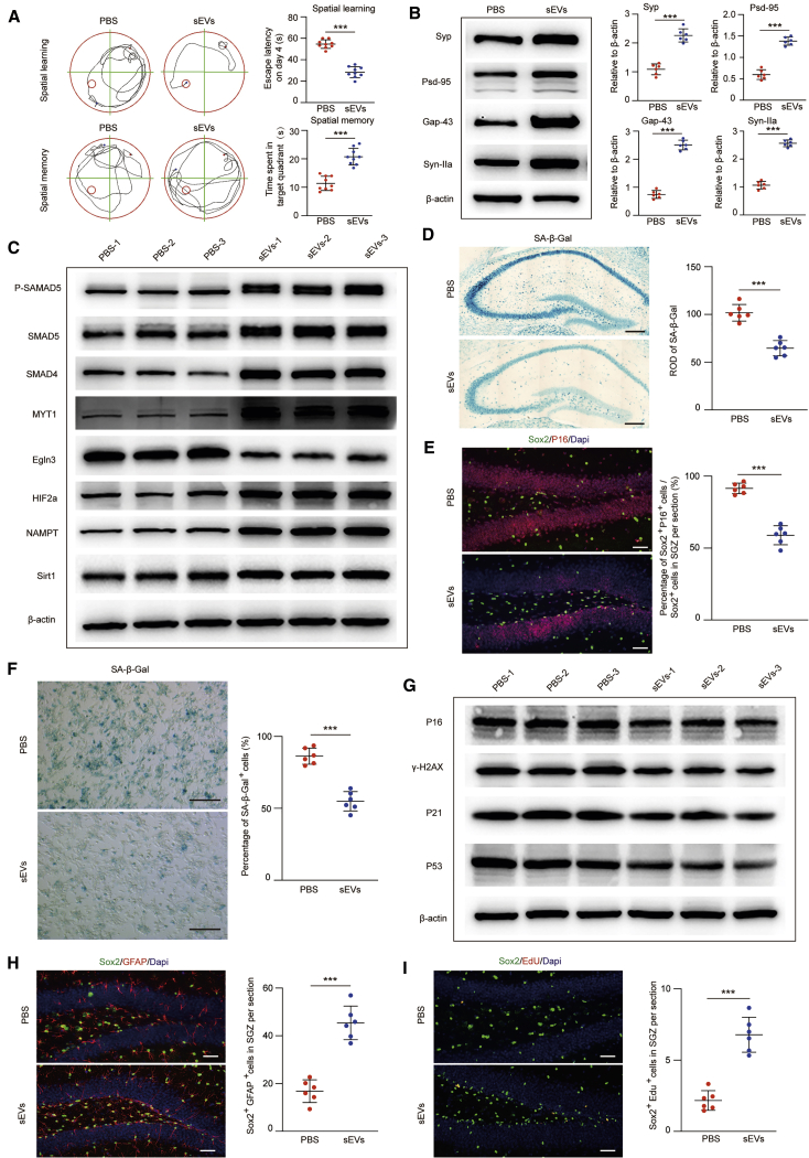
Tissue stem cell senescence leads to stem cell exhaustion, which results in tissue homeostasis imbalance and a decline in regeneration capacity. However, whether neural stem cell (NSC) senescence occurs and causes neurogenesis reduction during aging is unknown. In this study, mice at different ages were used to detect age-related hippocampal NSC (H-NSC) senescence, as well as the function and mechanism of embryonic stem cell-derived small extracellular vesicles (ESC-sEVs) in rejuvenating H-NSC senescence. We found a progressive cognitive impairment, as well as age-related H-NSC senescence, in mice. ESC-sEV treatment significantly alleviated H-NSC senescence, recovered compromised self-renewal and neurogenesis capacities, and reversed cognitive impairment. Transcriptome analysis revealed that myelin transcription factor 1 (MYT1) is downregulated in senescent H-NSCs but upregulated by ESC-sEV treatment. In addition, knockdown of MYT1 in young H-NSCs accelerated age-related phenotypes and impaired proliferation and differentiation capacities. Mechanistically, ESC-sEVs rejuvenated senescent H-NSCs partly by transferring SMAD family members 4 (SMAD4) and 5 (SMAD5) to activate MYT1, which downregulated egl-9 family hypoxia inducible factor 3 (Egln3), followed by activation of hypoxia inducible factor 2 subunit α (HIF-2α), nicotinamide phosphoribosyl transferase (NAMPT), and sirtuin 1 (Sirt1) successively. Taken together, our results indicated that H-NSC senescence caused cellular exhaustion, neurogenesis reduction, and cognitive impairment during aging, which can be reversed by ESC-sEVs. Thus, ESC-sEVs may be promising therapeutic candidates for age-related diseases.

Keywords: ESC-sEVs; MYT1; aging; hippocampal NSCs; senescence.


Figures

None
Graphical abstract
Figure 1
Figure 1
Cognitive Impairment, H-NSC Depletion, and Senescence in SAMP8 Mice during Aging (A) Spatial learning and memory abilities in mice at an age of 2, 6, and 12 months were tested by the MWM. n = 10 per group. ∗∗∗p < 0.001. (B) Western blot analysis and quantification of Syp, Gap-43, PSD-95, and Syn-IIa (relative to β-actin) in the hippocampus of mice at an age of 2, 6, and 12 months. n = 6 per group. ∗∗∗p < 0.001. (C) IF images for Sox2+ (green) and Nestin+ (red) cells and the number of Sox2+/Nestin+ double-stained cells in the SGZ of mice at an age of 2, 6, and 12 months. Scale bars, 50 μm. n = 6 per group. ∗∗∗p < 0.001. (D) IF images for Sox2+ (green) and GFAP+ (red) cells and the number of Sox2+/GFAP+ double-stained cells in the SGZ of mice at an age of 2, 6, and 12 months. Scale bars, 50 μm. n = 6 per group. ∗∗∗p < 0.001. (E) SA-β-gal staining of the hippocampus and quantification of hippocampal SA-β-gal activity in mice at an age of 2, 6, and 12 months. Scale bars, 250 μm. n = 6 per group. ∗∗∗p < 0.001. (F) IF images of p16INK4a+ (red)/Sox2+ (green) senescent H-NSCs and the quantification of Sox2+/p16INK4a+ double-stained cells of all Sox2+ cells in the SGZ of mice at an age of 2, 6, and 12 months. Scale bars, 50 μm. n = 6 per group. ∗∗∗p < 0.001. (G) Representative images of SA-β-gal staining and quantification of SA-β-gal+ cells from isolated H-NSCs from SAMP8 mice at an age of 2, 6, and 12 months. Scale bars, 100 μm. n = 6 per group. ∗∗∗p < 0.001. (H) Western blot analysis and quantification of p16INK4a, γ-H2AX, p21, and p53 in isolated neurospheres in SAMP8 mice at an age of 2, 6, and 12 months. n = 6 per group. ∗∗∗p < 0.001.
Figure 2
Figure 2
ESC-sEVs Reverse Cognitive Aging and Rejuvenate H-NSC Senescence in SAMP8 Mice (A) Spatial learning and memory abilities were tested by an MWM test in 6 month, 12 month-PBS, and 12 month-sEV mice. n = 10 per group. ∗∗∗p < 0.01. (B) Western blot analysis and quantification of Syp, Gap-43, PSD-95, and Syn-IIa in hippocampus of 6 month, 12 month-PBS, and 12 month-sEV mice. n = 6 per group. ∗∗∗p < 0.001. (C) SA-β-gal staining of the hippocampus and quantification of hippocampal SA-β-gal activity in 6 month, 12 month-PBS, and 12 month-sEV mice. Scale bars, 250 μm. n = 6 per group. ∗∗∗p < 0.001. (D) IF images of senescent H-NSCs (p16INK4a+, red; Sox2+, green) and the percentage of Sox2+/p16INK4a+ double-stained cells in whole Sox2+ cells in the SGZ of 6 month, 12 month-PBS, and 12 month-sEV mice. Scale bars, 50 μm. n = 6 per group. ∗∗∗p < 0.001. (E) SA-β-gal staining of isolated H-NSCs and the percentage of SA-β-gal+ cells in 6 month, 12 month-PBS, and 12 month-sEV mice. Scale bars, 100 μm. n = 6 per group. ∗∗∗p < 0.001. (F) Western blot analysis and quantification of p16INK4a, γ-H2AX, p21, and p53 in isolated neurospheres of 6 month, 12 month-PBS, and 12 month-sEV mice. n = 6 per group. ∗∗∗p < 0.001.
Figure 3
Figure 3
ESC-sEVs Reverse H-NSC Depletion and Promote Neurogenesis in the SGZ of SAMP8 Mice (A) IF images for hippocampal Sox2+ (green) and Nestin+ (red) cells and the number of Sox2+/Nestin+ double-stained cells in the SGZ of 6 month, 12 month-PBS, and 12 month-sEV mice. Scale bars, 50 μm. n = 6 per group. ∗∗∗p < 0.001. (B) IF images for hippocampal Sox2+ (green) and GFAP+ (red) cells and the number of Sox2+/GFAP+ double-stained cells in the SGZ of 6 month, 12 month-PBS, and 12 month-sEV mice. Scale bars, 50 μm. n = 6 per group. ∗∗∗p < 0.001. (C) IF images for proliferated H-NSCs (Sox2+, green; PCNA+, red) and the number of Sox2+/PCNA+ double-stained cells in the SGZ of 6 month, 12 month-PBS, and 12 month-sEV mice. Scale bars, 50 μm. n = 6 per group. ∗∗∗p < 0.001. (D) IF images for hippocampal DCX+ (green) and PCNA+ (red) cells and the number of DCX+/PCNA+ double-stained cells in the SGZ of 6 month, 12 month-PBS, and 12 month-sEV mice. Scale bars, 50 μm. n = 6 per group. ∗∗∗p < 0.001. (E) Neurosphere formation and quantification of neurosphere diameters in 6 month, 12 month-PBS, and 12 month-sEV mice. Scale bars, 300 μm. n = 6 per group. ∗p < 0.05. (F) IF images for EdU incorporation in isolated neurospheres and the percentage of EdU+/DAPI+ double-stained cells from total DAPI+ cells in 6 month, 12 month-PBS, and 12 month-sEV mice. Scale bars, 100 μm. n = 6 per group. ∗∗∗p < 0.001. (G) IF images for β-tubulin III+ (green) and GFAP+ cells and the percentage of β-tubulin III+ cells in whole cells in 6 month, 12 month-PBS, 12 month-ESC-sEV mice. Scale bars, 100 μm. n = 6 per group. ∗∗∗p < 0.001.
Figure 4
Figure 4
ESC-sEVs Upregulate MYT1 in H-NSCs and Ameliorate H-NSC Senescence (A) Representative images of SA-β-gal staining and the percentage of SA-β-gal+ cells in passage 2 (P2) H-NSCs, passage 10 H-NSCs treated with PBS (P10-PBS), and passage 10 H-NSCs treated with ESC-sEVs (P10-sEVs). Scale bars, 100 μm. n = 6 per group. ∗∗∗p < 0.001. (B) Western blot analysis and quantification of p16INK4a, γ-H2AX, p21, and p53 in H-NSCs of P2, P10-PBS, and P10-sEVs. n = 6 per group. ∗∗∗p < 0.001. (C) Neurosphere formation and quantification of neurosphere diameters in H-NSCs of P2, P10-PBS, and P10-sEVs. Scale bars, 300 μm. n = 6 per group. ∗p < 0.05. (D) IF images for EdU incorporation in neurospheres and the percentage of EdU+/DAPI+ double-stained cells in whole DAPI+ cells in H-NSCs of P2, P10-PBS, and P10-sEVs. Scale bars, 100 μm. n = 6 per group. ∗∗∗p < 0.001. (E) IF images for β-tubulin III+ (green) and GFAP+ cells and the percentage of β-tubulin III+ cells in whole cells in H-NSCs of P2, P10-PBS, and P10-sEVs. Scale bars, 100 μm. n = 6 per group. ∗∗∗p < 0.001. (F) RNA-seq data from H-NSCs of P2, P10-PBS, and P10-sEVs were analyzed by the “Calculate and draw custom Venn diagrams” online tool; 232 genes were co-upregulated and 415 genes were co-downregulated in P2 versus P10-PBS and P10-sEVs versus P10-PBS. n = 4 per group. (G) The relative expression level of MYT1 in H-NSCs of P2, P10-PBS, and P10-sEVs was detected by qRT-PCR. n = 6 per group. ∗∗∗p < 0.001. (H) Western blot analysis and quantification of MYT1 in H-NSCs of P2, P10-PBS, and P10-sEVs. n = 6 per group. ∗∗∗p < 0.001. (I) Western blot analysis and quantification of MYT1 in isolated neurospheres of 2 month, 6 month, and 12 month-PBS and 12 month-sEV mice. n = 6 per group. ∗∗∗p < 0.001.
Figure 5
Figure 5
Knockdown of MYT1 Accelerates H-NSC Senescence and Abolishes the Anti-aging Function of ESC-sEVs (A) The knockdown efficiency of MYT1 shRNAs in H-NSCs was detected by qRT-PCR. n = 3 per group. ∗∗p < 0.01, ∗∗∗p < 0.001. (B) Western blot analysis and quantification of MYT1 in H-NSCs showing the knockdown efficiency of MYT1 shRNAs. n = 3 per group. ∗∗p < 0.01, ∗∗∗p < 0.001. (C) SA-β-gal staining and the percentage of SA-β-gal+ cells in sh-NC NSCs treated with PBS or ESC-sEVs (sh-NC-PBS or sh-NC-sEVs) and sh-MYT1 NSCs treated with PBS or ESC-sEVs (sh-MYT1-PBS or sh-MYT1-sEVs). Scale bars, 100 μm. n = 6 per group. ∗∗∗p < 0.001. (D) Western blot analysis and quantification of p16INK4a, γ-H2AX, p21, and p53 in H-NSCs of sh-NC-PBS, sh-NC-sEVs, sh-MYT1-PBS, and sh-MYT1-sEVs. n = 6 per group. ∗∗∗p < 0.001. (E) IF images for GFP-labeled neurospheres and the diameter of neurospheres in H-NSCs of sh-NC-PBS, sh-NC-sEVs, sh-MYT1-PBS, and sh-MYT1-sEVs. Scale bars, 200 μm. n = 6 per group. ∗p < 0.05. (F) IF images for EdU incorporation in neurospheres and the percentage of EdU+/DAPI+ double-stained cells in all DAPI+ cells in sh-NC-PBS, sh-NC-sEVs, sh-MYT1-PBS, and sh-MYT1-sEVs. Scale bars, 100 μm. n = 6 per group. ∗∗∗p < 0.001. (G) IF images for β-tubulin III+ and the percentage of β-tubulin III+ cells in whole cells in H-NSCs of sh-NC-PBS, sh-NC-sEVs, sh-MYT1-PBS, and sh-MYT1-sEVs. Scale bars, 100 μm. n = 6 per group. ∗∗∗p < 0.001.
Figure 6
Figure 6
ESC-sEVs Rejuvenate Senescent H-NSCs by Transferring SMADs to Activate MYT1, Inhibit Egln3, and Upregulate HIF-2α, NAMPT, and Sirt1 Successively (A) Western blot analysis and quantification of MYT1, Egln3, HIF-2α, NAMPT, and Sirt1 in H-NSCs of sh-NC-PBS, sh-NC-sEVs, sh-MYT1-PBS, and sh-MYT1-sEVs. n = 6 per group. ∗∗∗p < 0.001. (B) Comparison of proteins in ESC-sEVs identified by LC-MS/MS with the EV proteome database Vesiclepedia. (C) GO analysis of proteins in ESC-sEVs showed mostly that proteins belonged to extracellular vesicles. (D) Western blot analysis of SMAD4, SMAD5, and TSG101 in ESCs, ESC-sEVs, and PBS. (E) Western blot analysis and quantification of p-SMAD5, SMAD5, SMAD4, and MYT1 in H-NSCs treated with PBS and ESC-sEVs. (F) Western blot analysis and quantification of p-SMAD5, SMAD5, SMAD4, and MYT1 in H-NSCs, H-NSCs treated with 25 ng/mL BMP4, H-NSCs treated with 200 nM LDN-193189, and H-NSCs treated with LDN-193189 and BMP4. n = 3 per group. ∗∗p < 0.01, ∗∗∗p < 0.001. (G) Western blot analysis and quantification of p-SMAD5, SMAD5, SMAD4, MYT1, Egln3, HIF-2α, NAMPT, and Sirt1 in isolated neurospheres of 2 month, 6 month, and 12 month-PBS and 12 month-sEV mice. n = 6 per group. ∗∗∗p < 0.001.
Figure 7
Figure 7
ESC-sEVs Reverse Cognitive Aging and Rejuvenate H-NSC Senescence in C57BL/6 Mice (A) Spatial learning and memory abilities were tested by an MWM test in 20 month-PBS and 20 month-sEV mice. n = 9 per group. ∗∗∗p < 0.001. (B) Western blot analysis and quantification of Syp, Gap-43, PSD-95, and Syn-IIa in hippocampus of 20 month-PBS and 20 month-sEV mice. n = 6 per group. ∗∗∗p < 0.001. (C) Western blot analysis of p-SMAD5, SMAD5, SMAD4, MYT1, Egln3, HIF-2α, NAMPT, and Sirt1 in isolated neurospheres of 20 month-PBS and 20 month-sEV mice. n = 3 per group. (D) SA-β-gal staining of hippocampus and quantification of hippocampal SA-β-gal activity in 20 month-PBS and 20 month-sEV mice. Scale bars, 250 μm. n = 6 per group. ∗∗∗p < 0.001. (E) IF images for senescent H-NSCs (p16INK4a+, red; Sox2+, green) and the percentage of Sox2+/p16INK4a+ double-stained cells in whole Sox2+ cells in the SGZ of 20 month-PBS and 20 month-sEV mice. Scale bars, 50 μm. n = 6 per group. ∗∗∗p < 0.001. (F) SA-β-gal staining of isolated H-NSCs and the percentage of SA-β-gal+ cells in 20 month-PBS and 20 month-sEV mice. Scale bars, 100 μm. n = 3 per group. ∗∗∗p < 0.001. (G) Western blot analysis of p16INK4a, γ-H2AX, p21, and p53 in isolated neurospheres of 20 month-PBS and 20 month-sEV mice. n = 3 per group. (H) IF images for hippocampal Sox2+ (green) and GFAP+ (red) cells and the number of Sox2+/GFAP+ double-stained cells in the SGZ of 20 month-PBS and 20 month-sEV mice. Scale bars, 50 μm. n = 6 per group. ∗∗∗p < 0.001. (I) IF images for proliferating H-NSCs (Sox2+, green; EdU+, red) and the number of Sox2+/EdU+ double-stained cells in the SGZ of 20 month-PBS and 20 month-sEV mice. Scale bars, 50 μm. n = 6 per group. ∗∗∗p < 0.001.

Similar articles

Cited by

References

    1. Nguyen H., Zarriello S., Coats A., Nelson C., Kingsbury C., Gorsky A., Rajani M., Neal E.G., Borlongan C.V. Stem cell therapy for neurological disorders: a focus on aging. Neurobiol. Dis. 2019;126:85–104. - PMC - PubMed
    1. Capilla-Gonzalez V., Herranz-Pérez V., García-Verdugo J.M. The aged brain: genesis and fate of residual progenitor cells in the subventricular zone. Front. Cell. Neurosci. 2015;9:365. - PMC - PubMed
    1. Encinas J.M., Michurina T.V., Peunova N., Park J.H., Tordo J., Peterson D.A., Fishell G., Koulakov A., Enikolopov G. Division-coupled astrocytic differentiation and age-related depletion of neural stem cells in the adult hippocampus. Cell Stem Cell. 2011;8:566–579. - PMC - PubMed
    1. Bouab M., Paliouras G.N., Aumont A., Forest-Bérard K., Fernandes K.J. Aging of the subventricular zone neural stem cell niche: evidence for quiescence-associated changes between early and mid-adulthood. Neuroscience. 2011;173:135–149. - PubMed
    1. Childs B.G., Durik M., Baker D.J., van Deursen J.M. Cellular senescence in aging and age-related disease: from mechanisms to therapy. Nat. Med. 2015;21:1424–1435. - PMC - PubMed

Publication types

MeSH terms

LinkOut - more resources