. 2020 Feb 10;37(2):200-215.e5.
doi: 10.1016/j.ccell.2020.01.001.

Direct Phosphorylation and Stabilization of MYC by Aurora B Kinase Promote T-cell Leukemogenesis

Affiliations

Direct Phosphorylation and Stabilization of MYC by Aurora B Kinase Promote T-cell Leukemogenesis

Jue Jiang et al. Cancer Cell. .

Abstract

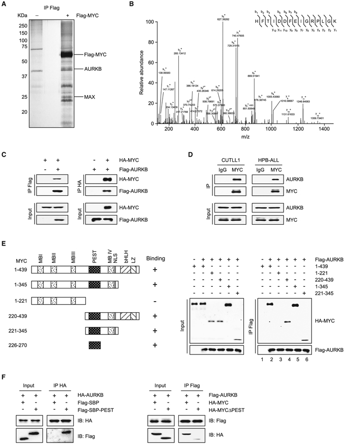
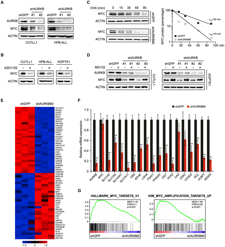
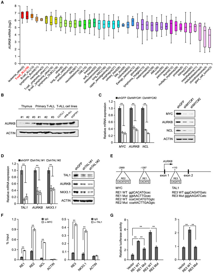
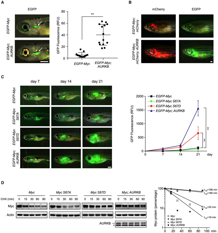
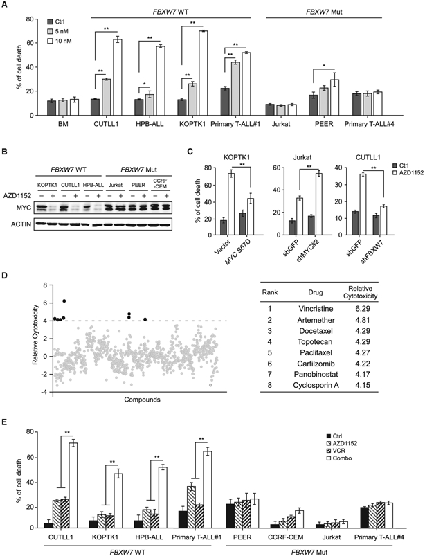
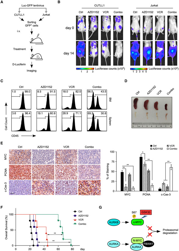
Deregulation of MYC plays an essential role in T cell acute lymphoblastic leukemia (T-ALL), yet the mechanisms underlying its deregulation remain elusive. Herein, we identify a molecular mechanism responsible for reciprocal activation between Aurora B kinase (AURKB) and MYC. AURKB directly phosphorylates MYC at serine 67, counteracting GSK3β-directed threonine 58 phosphorylation and subsequent FBXW7-mediated proteasomal degradation. Stabilized MYC, in concert with T cell acute lymphoblastic leukemia 1 (TAL1), directly activates AURKB transcription, constituting a positive feedforward loop that reinforces MYC-regulated oncogenic programs. Therefore, inhibitors of AURKB induce prominent MYC degradation concomitant with robust leukemia cell death. These findings reveal an AURKB-MYC regulatory circuit that underlies T cell leukemogenesis, and provide a rationale for therapeutic targeting of oncogenic MYC via AURKB inhibition.

Keywords: Aurora B kinase; FBXW7; MYC; T-ALL; patient-derived xenograft; phosphorylation; protein stability; zebrafish T-ALL model.


Conflict of interest statement

Declaration of Interests The authors declare no competing interests.

Figures

Figure 1.
Figure 1.. MYC Interacts with AURKB
(A) Purification of MYC and its interacting partners. Total protein extract from Jurkat cells expressing Flag-tagged MYC or vector was subjected to immunoprecipitation using anti-Flag beads. Eluted proteins were resolved by SDS-PAGE and visualized by silver staining. (B) Liquid chromatography-tandem mass spectrometry analysis of the Flag-MYC-associated peptides corresponding to AURKB. (C) Lysates of 293T cells overexpressing Flag-AURKB and/or HA-MYC were subjected to reciprocal co-immunoprecipitation (co-IP) to detect protein interaction. (D) CUTLL1 and HPB-ALL cell lysates were subjected to co-IP and immunoblot to detect endogenous MYC and AURKB interaction. (E) Schematic presentation of various human MYC truncations used in AURKB-binding assays (left). Lysates from 293T cells overexpressing Flag-AURKB and/or various HA-MYC truncations were subjected to co-IP and immunoblot (right). (F) Co-IP of HA-AURKB and Flag-tagged streptavidin-binding peptide (SBP)-fused MYC PEST domain (left), or Flag-AURKB and HA-MYCΔPEST (right) from lysates of 293T cells overexpressing respective tagged proteins. See also Table S1.
Figure 2.
Figure 2.. AURKB Inhibition Attenuates the MYC Protein Accumulation and Transcriptional Activity
(A) AURKB was depleted by specific shRNAs in T-ALL cells as indicated. AURKB and MYC proteins were analyzed by immunoblot, with ACTIN as a loading control. shGFP, control shRNA targeting GFP. (B) Immunoblots of MYC and ACTIN in T-ALL cells treated with 10 nM AZD1152 for 24 h. (C) Time-course analysis of MYC protein levels in AURKB-depleted CUTLL1 cells. MYC proteins were quantified and plotted on the right. (D) AURKB-depleted CUTLL1 and KOPTK1 cells were treated with MG132 (10 μM) for 6 h before harvest. AURKB and MYC were analyzed by immunoblot, with ACTIN as a loading control. (E) A heatmap depicting top 30 down- and upregulated genes upon AURKB depletion in CUTLL1 cells (Q < 0.05). (F) qRT-PCR analysis of representative MYC-induced target genes in CUTLL1 cells upon AURKB depletion. Data shown represent the means (±SD) of biological triplicates. *p < 0.05, **p < 0.01, unpaired two-tailed Student’s t test. (G) Gene set enrichment analysis of MYC target gene sets in the expression profiles of CUTLL1 cells expressing AURKB shRNA (http://software.broadinstitute.org/gsea). See also Figure S1.
Figure 3.
Figure 3.. AURKB Enhances MYC Protein Stability through the S67 Phosphorylation
(A) Time-course analysis of MYC p-T58 by immunoblot upon AZD1152 (50 nM) treatment in CUTLL1 and HPB-ALL cells, with ACTIN as a loading control. (B) Protein interaction between GSK3β and MYC in the presence of AURKB. pCDH-Flag-GSK3β (1 μg), pCDH-HA-MYC (1 μg), and/or increasing doses of pCDH-AURKB (0.5,1, and 2 μg) were transfected into 293T cells for 48 h as indicated. Cell lysates were then subjected to co-IP and immunoblot. The star (*) designates immunoglobulin G heavy chain. (C) Time-course analysis of HA-MYC levels in 293T cells expressing ectopic HA-MYC, Flag-AURKB WT, or mutant (K106R) as indicated. MYC proteins were quantified and plotted on the right. Data shown are means (±SD) of three independent experiments. (D) Peptide sequence alignment of MYC (amino acids 58–74) in various species. The S67 site resides in the AURKB phosphorylation consensus motif. (E) Time-course analysisofHA-tagged MYC(WT ormutants) byimmunoblot in 293T cells(left). MYC proteinswerequantified and plotted ontheright. Datashown are means (±SD) of three independent experiments. (F) Co-IP of Flag-GSK3β and HA-MYC (WT, S67D, or S67A) from lysates of 293T cells overexpressing respective tagged proteins. (G) In vitro kinase analysis of recombinant GST-MYC. Active human AURKB proteins were incubated with GST-MYC for kinase reaction. Phosphorylated proteins were separated by SDS-PAGE and visualized by autoradiography. Loading controls were shown as Coomassie blue staining in the bottom panels. (H) Detection of endogenous MYC p-S67. CUTLL1 and primary T-ALL cells were treated with Nocodazole(Noco, 1 μg/mL) for 4 h or AZD1152 (10 nM) for 24 h as indicated. Noco-treated cell lysates were treated with calf-intestinal alkaline phosphatase (CIP) for 30 min before immunoblot. Histone 3 p-S10 (p-S10 H3) levels were shown as a positive control reflecting AURKB activity. See also Figure S2.
Figure 4.
Figure 4.. MYC and TAL1 Activate AURKB Transcription
(A) AURKB mRNA expression was analyzed among 1,457 human cancer cell lines in CCLE database (https://portals.broadinstitute.org/ccle). The distributions of AURKB mRNA expression are presented as log2 median-centered intensity and shown in box-and-whisker plots, which depict the first and third quartiles, with the median shown as a solid line inside the box and whiskers extending to 1.5 interquartile range from first and third quartiles. (B) AURKB proteins were analyzed by immunoblot in normal thymuses from healthy donors, primary T-ALL samples and T-ALL cell lines, respectively. (C) AURKB mRNA and protein were analyzed in MYC-depleted CUTLL1 cells by qRT-PCR and immunoblot. MYC target gene NCL was used as a positive control. (D) AURKB mRNA and protein were analyzed in TAL1-depleted Jurkat cells by qRT-PCR and immunoblot. TAL1 target gene NKX3.1 was used as a positive control. (E) Schematic presentation of MYC and TAL1 binding sites on the AURKB locus. MYC response elements, RE1 and RE2; TAL1 responsive element, RE3. Consensus sequence mutations are shown as RE Mut. (F) ChIP analysis of MYC binding to the AURKB locus in CUTLL1 cells (left) orTAL1 binding in Jurkat cells (right). Binding signals to NCL and NKX3.1 are shown as positive controls, ACTIN as a negative control. (G) Luciferase reporter activities were assessed using MYC3×RE constructs in the presence of exogenous MYC in 293T cells (left) or when co-expressing TAL1 3×RE reporter constructs with TAL1 (right). Data shown represent the means (±SD) of three biological replicates, **p < 0.01; significance was determined by unpaired two-tailed Student’s t test (F) or one-way ANOVA test followed by Tukey’s correction (C, D, and G). See also Figure S3.
Figure 5.
Figure 5.. AURKB Promotes Murine Myc-Induced T-ALL
(A) Zebrafish embryos were injected respectively with the rag2:EGFP-Myc construct alone or in combination with rag2:AURKB. Representative images of GFP fluorescent microscopy analysis at 8 dpf are shown on the left. GFP fluorescence from the thymuses of EGFP-Myc (n = 13) and EGFP-Myc;AURKB fish (n = 14) were qualified and plotted on the right. Scale bar, 1 mm. Data are means ± SD, **p < 0.01, unpaired two-tailed Student’s t test. (B) Representative images of GFP or mCherry dissemination at 80 dpf in EGFP-Myc;mCherty (n = 10) and EGFP-Myc;mCherry;AURKB (n = 12) transgenic fish receiving heat shock treatments. Scale bar, 1 mm. (C) Representative GFP images of zebrafish injected with indicated constructs (left). Scale bar, 1 mm. Quantifications of the GFP fluorescence are shown on the right (n = 6 in each group). Data are mean ± SD, **p < 0.01, two-way ANOVA test followed by Tukey’s correction. (D) Time-course analysis of exogenous murine Myc from zebrafish expressing various transgenes as indicated. The arrow denotes human AURKB transgene expression and the bands below (*) are endogenous zebrafish Aurkb. Zebrafish Actin was used as a loading control (left). Myc protein levels at each point were quantified and plotted on the right. See also Figure S4.
Figure 6.
Figure 6.. AURKB Inhibition Induces Apoptosis in T-ALL Cells Expressing WT FBXW7
(A) Analysis of apoptotic cell death by Annexin V/PI staining in normal bone marrow cells and a panel of T-ALL cell lines, harboring WT or mutant FBXW7, treated with AZD1152 for 48 h. (B) Immunoblots of MYC in 10 nM AZD1152-treated T-ALL cells. (C) Cell death analysis in MYC S67D expressing KOPTK1 cells (left), MYC-depleted Jurkat cells (middle), or FBXW7-depleted CUTLL1 cells (right), which were subjected to 5, 25, or 5 nM AZD1152 treatment, respectively. (D) High-throughput screening of US Food and Drug Administration-approved drugs in KOPTK1 cells to identify small molecules synergistic with AZD1152. Top hits are listed on the right. (E) Assessment of cell death in T-ALL cells treated with AZD1152 (5 nM) and/or vincristine (1 nM) for 48 h. Combo, combination treatment. Data shown represent the means (±SD) of three biological replicates, *p < 0.05, **p < 0.01; significance was determined by one-way (A and E) or two-way (C) ANOVA test followed by Tukey’s correction. See also Figures S5 and S6 and Table S2.
Figure 7.
Figure 7.. AZD1152 and Vincristine Synergistically Inhibit Xenograft Tumor Growth
(A) Schematic representation of in vivo imaging of T-ALL xenografts. CUTLL1 (FBXW7 WT) or Jurkat (FBXW7 Mut) cells, expressing both luciferase and GFP markers (Luc-GFP), were injected into NPG mice and subjected to treatments, followed by in vivo bioimaging to assess therapeutic responses. (B) Representative images of tumor burden assessed by bioimaging in mice xenografted with CUTLL1 or Jurkat cells upon single or combination treatments (n = 3 per group). Drug administrations started at day 0. (C) Anti-leukemia effects of drug combination in a patient-derived xenograft (PDX). Human CD45+ cells from bone marrow (BM) and spleen were analyzed by flow cytometry. (D) Representative spleen and bone images of mice in each group at day 25 after engraftment. (E) Representative immunohistological images of MYC, proliferating cell nuclear antigen (PCNA), and cleaved caspase-3 (c-Cas-3) in the spleen sections from mice receiving indicated treatments. Scale bar, 50 μm. Quantifications of immunohistochemistry are shown on the right. Data are mean ± SD, *p < 0.05, **p < 0.01, one-way ANOVA test followed by Tukey’s correction. (F) Kaplan-Meier survival curves of T-ALL PDX treated with AZD1152 and/or vincristine (n = 5 in each group). Significance was determined by logrank test, **p < 0.01. (G) Model depicting the regulation of (c-)MYC or N-MYC by Aurora kinases through kinase-dependent or independent mechanism. See text for more details.

Similar articles

Cited by

References

    1. Archambault V, and Carmena M (2012). Polo-like kinase-activating kinases: Aurora A, Aurora B and what else? Cell Cycle 11, 1490–1495. - PMC - PubMed
    1. Bachireddy P, Bendapudi PK, and Felsher DW (2005). Getting at MYC through RAS. Clin. Cancer Res 11, 4278–4281. - PubMed
    1. Barretina J, Caponigro G, Stransky N, Venkatesan K, Margolin AA, Kim S, Wilson CJ, Lehar J, Kryukov GV, Sonkin D, et al. (2012). The Cancer Cell Line Encyclopedia enables predictive modelling of anticancerdrug sensitivity. Nature 483, 603–607. - PMC - PubMed
    1. Bonnet M, Loosveld M, Montpellier B, Navarro J-M, Quilichini B, Picard, , Di Cristofaro J, Bagnis C, Fossat C, Hernandez L, et al. (2011). Posttranscriptional deregulation of MYC via PTEN constitutes a majoralternative pathway of MYC activation in T-cell acute lymphoblastic leukemia. Blood 117, 6650–6659. - PubMed
    1. Chen H, Liu H, and Qing G (2018). Targeting oncogenic Myc as a strategy for cancer treatment. Signal. Transduct. Target Ther 3, 5. - PMC - PubMed

Publication types

MeSH terms