. 2020 Apr 17;48(7):3935-3948.
doi: 10.1093/nar/gkaa097.

Oplr16 serves as a novel chromatin factor to control stem cell fate by modulating pluripotency-specific chromosomal looping and TET2-mediated DNA demethylation

Affiliations

Oplr16 serves as a novel chromatin factor to control stem cell fate by modulating pluripotency-specific chromosomal looping and TET2-mediated DNA demethylation

Lin Jia et al. Nucleic Acids Res. .

Abstract

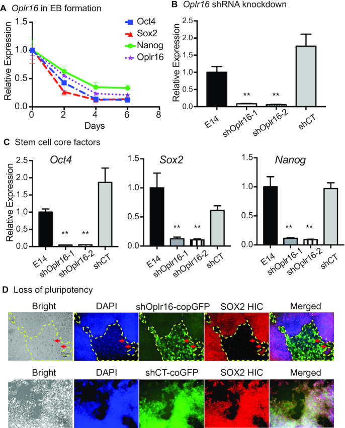
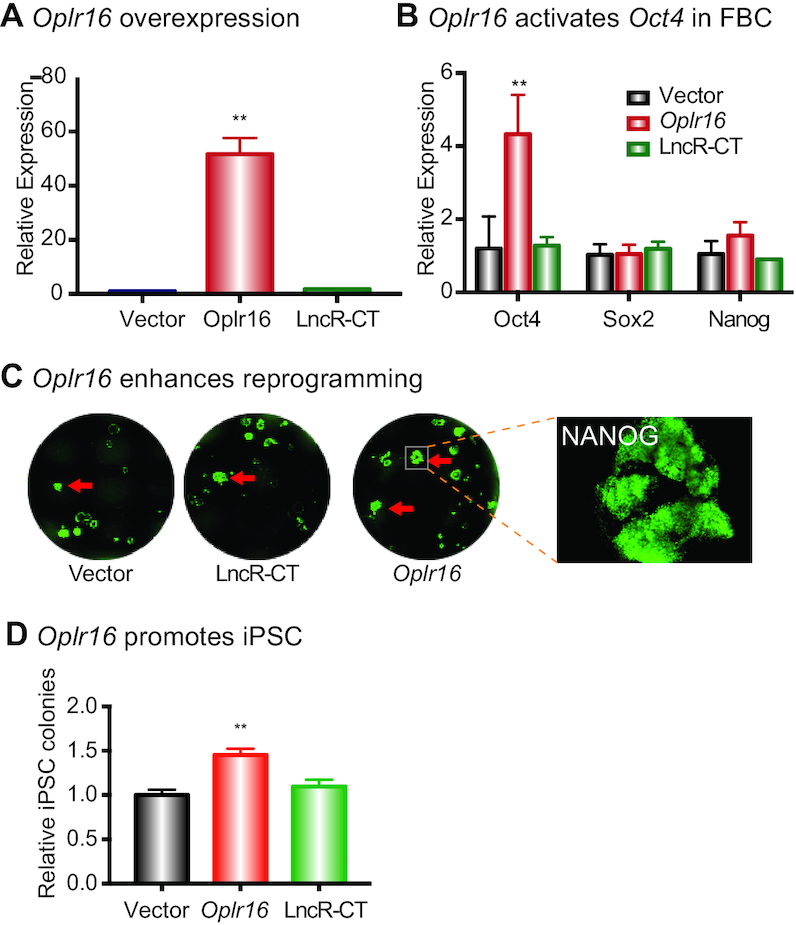
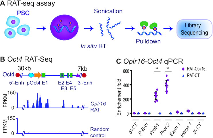
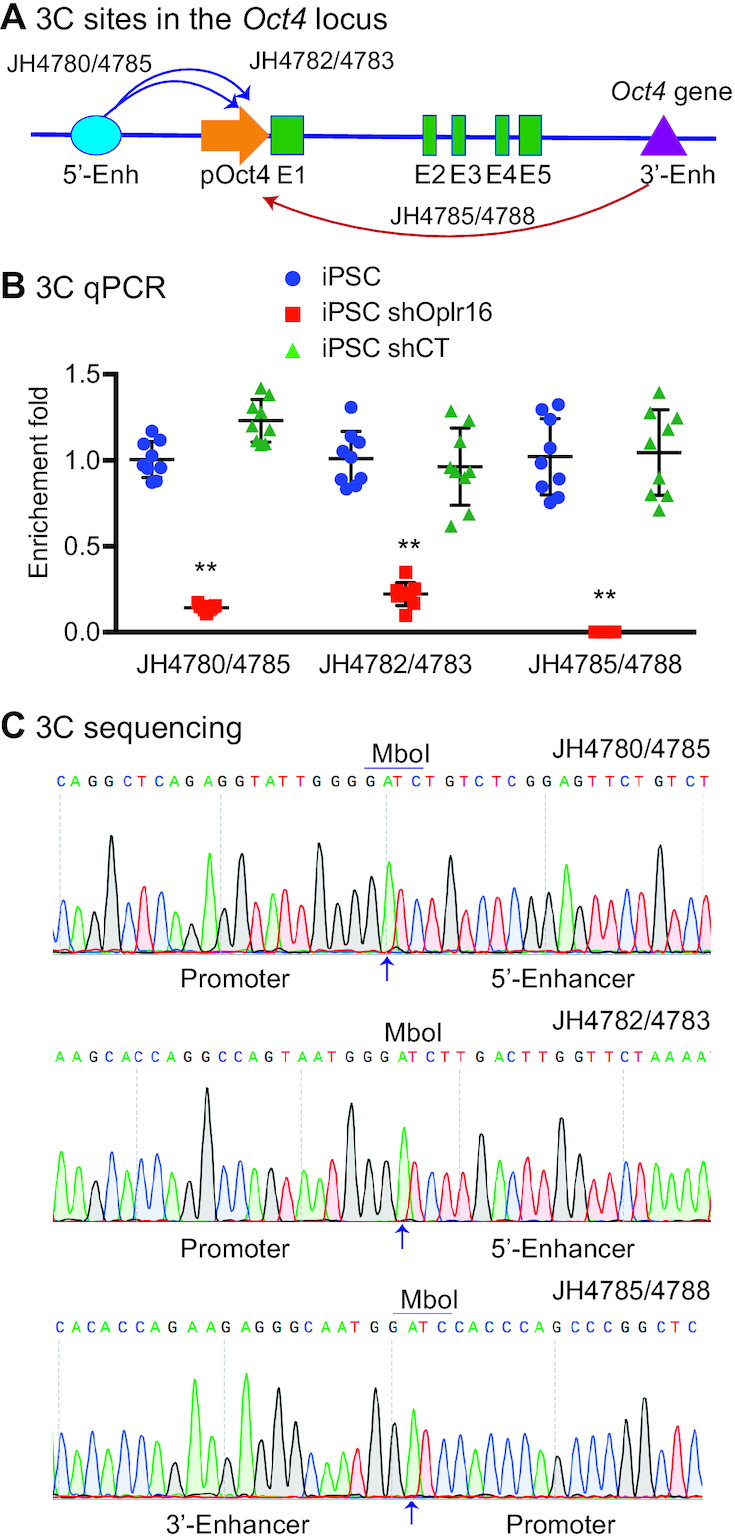
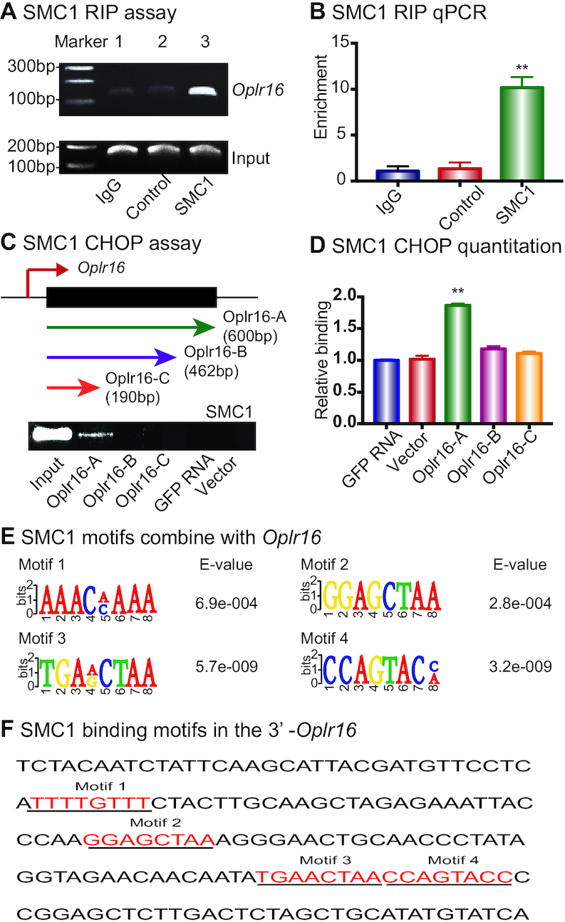
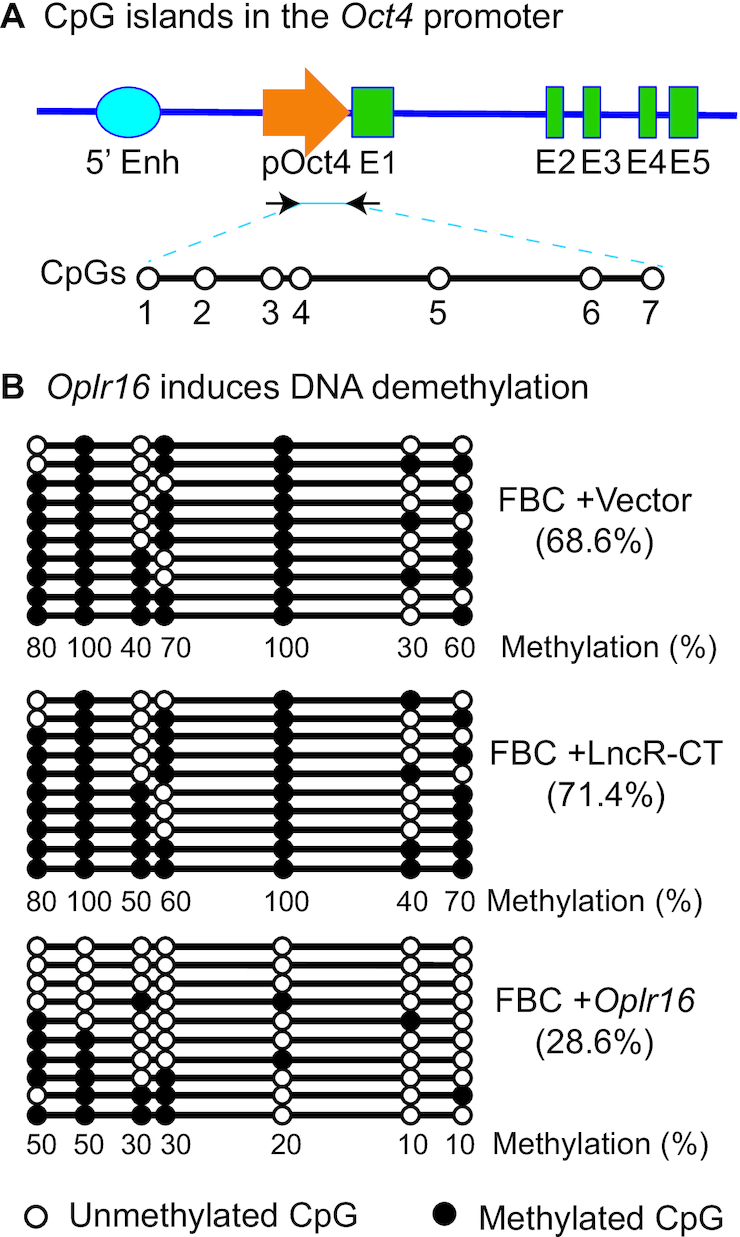
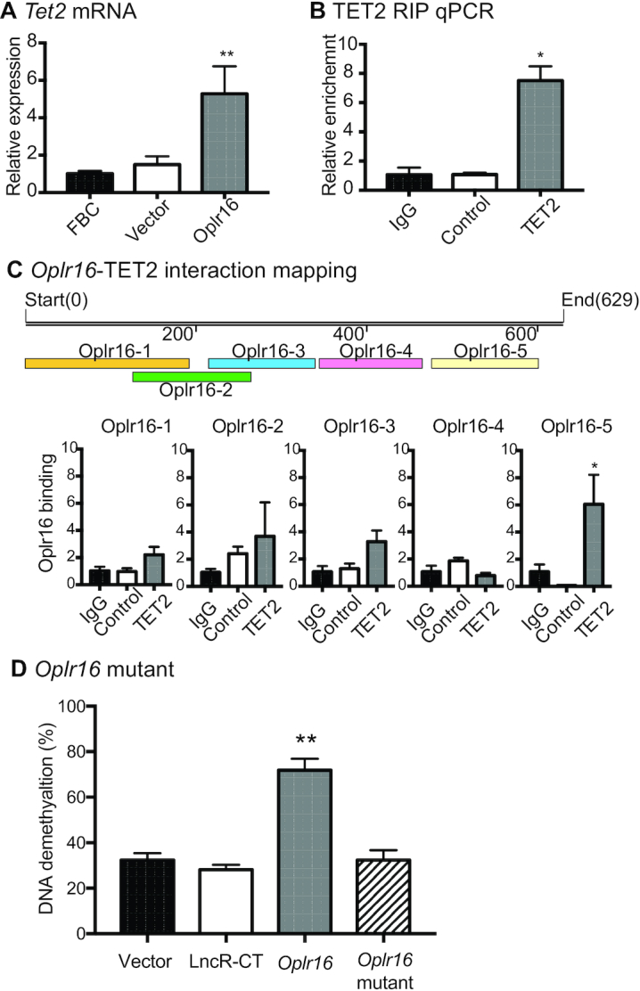
Formation of a pluripotency-specific chromatin network is a critical event in reprogramming somatic cells into pluripotent status. To characterize the regulatory components in this process, we used 'chromatin RNA in situ reverse transcription sequencing' (CRIST-seq) to profile RNA components that interact with the pluripotency master gene Oct4. Using this approach, we identified a novel nuclear lncRNA Oplr16 that was closely involved in the initiation of reprogramming. Oplr16 not only interacted with the Oct4 promoter and regulated its activity, but it was also specifically activated during reprogramming to pluripotency. Active expression of Oplr16 was required for optimal maintenance of pluripotency in embryonic stem cells. Oplr16 was also able to enhance reprogramming of fibroblasts into pluripotent cells. RNA reverse transcription-associated trap sequencing (RAT-seq) indicated that Oplr16 interacted with multiple target genes related to stem cell self-renewal. Of note, Oplr16 utilized its 3'-fragment to recruit the chromatin factor SMC1 to orchestrate pluripotency-specific intrachromosomal looping. After binding to the Oct4 promoter, Oplr16 recruited TET2 to induce DNA demethylation and activate Oct4 in fibroblasts, leading to enhanced reprogramming. These data suggest that Oplr16 may act as a pivotal chromatin factor to control stem cell fate by modulating chromatin architecture and DNA demethylation.


Figures

Figure 1.
Figure 1.
Identification of Oplr16 as a pluripotent lncRNA. (A) Identification of Oplr16 as a pluripotency-associated lncRNA by integrating RNA-seq and CRIST-seq. RNA-seq was used to identify RNAs that are differentially expressed between fibroblasts and iPSCs collected in the process of reprogramming. In CRIST-seq, the Oct4 promoter was targeted by the expression of the Cas9 Oct4-gRNA. LncRNAs interacting with Oct4 promoter were reverse transcribed in situ into cDNA with biotin-dCTP. After immunoprecipitation, biotin-cDNAs were purified by streptavidin beads for Illumina sequencing. Both sets of databases were integrated to identify the pluripotency-associated lncRNA Oplr16. (B) Reactivation of Oplr16 in reprogramming. Fibroblasts were transfected with a Oct4-Sox2-Kilf4-c-Myc (OSKM) lentivirus. Cells were collected at various stages of reprogramming and expression of Oplr16 was measured by qPCR. E14: mouse embryonic pluripotent stem cell line used as a positive control; iPSC: induced pluripotent stem cells; non-iPSC: un-reprogrammed cells that express four OSKM factors, but fail to complete reprogramming; FBC: fibroblasts. β-Actin was used as the PCR control. ** P < 0.01 as compared with FBC and URC. (C) Oplr16 is predominantly located in the nucleus. Cytoplasmic (cyto) and nuclear RNAs were isolated and reverse transcribed into cDNAs. Location of Oplr16 was determined by PCR. β-Actin: cytoplasm control. U6: nuclear control. (D) Confirmation of nuclear location of Oplr16 by RNA-FISH. Digoxygenin labeled Oplr16 probe was prepared and hybridized in iPSCs and fibroblasts (FBC). DAPI: nuclear control.
Figure 2.
Figure 2.
Oplr16 is required for the maintenance of stem cell pluripotency. (A) Expression of Oplr16 decreased during embryoid body differentiation. iPSCs were collected at Days 2, 4, 6 after EB formation and qPCR was used to quantitate the expression of Oplr16 in relation to that of stem cell core factor genes Oct4, Sox2 and Nanog. (B) Knockdown of Oplr16 by shRNAs in E14 cells. shOplr16-1 and shOplr16-2: shRNAs that target Oplr16; shCT: random shRNA control. ** P < 0.01 as compared with iPSC and shCT. (C) Downregulation of stem cell core factor genes Oct4, Sox2 and Nanog by Oplr16 knockdown. After transfection with shOplr16-1 and shOplr16-2 lentiviruses, cells were collected for quantitative PCR. ** P < 0.01 as compared with iPSC and shCT. (D) Knockdown of Oplr16 induces loss of pluripotency. Oplr16 was knocked down by shOplr16 lentiviruses in E14 cells. The shCT lentiviruses were used as control. Lentivirus-transfected cells were tracked by copGFP (green). The pluripotency was examined by histoimmunochemical (HIC) staining of stem cell marker SOX2 (red). ShOplr16-transfected E14 cells showed the loss of pluripotent marker SOX2. Cell morphology was also changed. As the control, the shCT treatment did not alter stem cell pluripotency.
Figure 3.
Figure 3.
Oplr16 promotes Reprogramming. (A) Overexpression of Oplr16 in fibroblasts. Fibroblasts were transfected by Oplr16 lentiviruses and control lentiviruses. After selection by puromycin, stable cells were collected for qPCR quantitation. (B) Oplr16 activated the endogenous Oct4 in transfected fibroblasts. However, the expression of Sox2 and Nanog show no significant change as compared with Vector and LncR-CT controls. (C) Oplr16 enhanced reprogramming. DOX-inducible MEF cells were transfected with lentiviruses carrying the empty vector, random lncRNA control, and Oplr16. After 10 days of doxycycline (DOX) induction, iPSC colonies were immunostained using an antibody against pluripotency maker NANOG (green). (D) Oplr16 promoted iPSC formation. After DOX induction, the number of NANOG positive iPSC colonies was quantitated. ** P < 0.01 as compared with the controls.
Figure 4.
Figure 4.
Oplr16 binds to the Oct4 promoter. (A) Schematic diagram of the RNA reverse transcription-associated trap sequencing (RAT-seq) assay. Oplr16 lncRNA was in situ reverse transcribed using three Oplr16-specific complementary primers with biotin-dCTP. The random primers were used as the negative control (RAT-CT). After nuclear lysis, the bitoin-Oplr16 cDNA chromatin complex was isolated by streptavidin beads and the Oplr16-interacting target DNAs were isolated for Illumina library sequencing. (B) The IGV analysis of Oplr16 binding signals in the Oct4 locus. 5′-Enh, 3′-Enh: the Oct4 5′- and 3′-enhancers; pOct4: Oct4 promoter; E1-E5: Oct4 exons. (C) Quantitative PCR mapping of Oplr16 binding in the Oct4 locus. The RAT pulldown complex was used to map the Oplr16 binding. 5′-CT, 3′-CT: the RAT control sites in the Oct4 locus. Note the enrichment of the Oplr16 binding signals in the promoter region (Prot-1 and Prot-2) (N = 9, ** P < 0.001 as compared with the RAT control).
Figure 5.
Figure 5.
Oplr16 is essential for the maintenance of intrachromosomal looping in the Oct4 locus. (A) The PCR sites in the Oct4 locus. Intrachromosomal interactions were quantitated by 3C (chromatin conformation capture) qPCR. 5′-Enh: 5′-enhancers; pOct4: Oct4 promoter; E1-E5: Exons; 3′-Enh: 3′-Enhancer. Arrows: intrachromosomal interactions. (B) Knockdown of Oplr16 abolished intrachromosomal interaction loops. The 3C interaction was detected by qPCR. For comparison, the relative 3C interaction was calculated by setting the 5′- and 3′- controls as 1 (N = 9, ** P < 0.001 as compared with the shCT and iPSC controls). (C) Sequencing of the Oct4 intrachromosomal loop products. Arrows: the 3C ligation product containing the MboI site that is flanked by the promoter and the enhancer sequences.
Figure 6.
Figure 6.
Oplr16 interacts with chromatin factor SMC1. (A) Interaction of Oplr16 with SMC1 by RNA-chromatin immunoprecipitation (RIP). The SMC1–lncRNA chromatin complex was immunoprecipitated with an antibody against SMC1. The immunoprecipitated RNAs were reverse transcribed and the SMC1-interacting Oplr16 was measured by PCR. IgG was used as the antibody control. Input: aliquot DNAs collected during the RIP assay. (B) Quantitative RIP PCR of the Oplr16-SMC1 interaction. (C) Identification of the SMC1 binding fragment in Oplr16. The chromatin oligo affinity precipitation (CHOP) assay was used to determine the specific fragment of Oplr16 that interacts with SMC1. Top panel: Schematic diagram of CHOP mapping. Three different sizes of biotin-labeled Oplr16 fragments were synthesized and bound to streptavidin agarose beads. Recombinant SMC1 proteins were incubated with biotin-Oplr16 streptavidin beads. After elution, the Oplr16-interacted SMC1 proteins were analyzed by Western immunoblotting. Only the full-length Oplr16 showed the interaction with SMC1 (Oplr16-A). GFP RNA was used as the negative control. Input: SMC1 protein. (D) Quantitation of the interaction of SMC1 with Oplr16 fragments. (E) Consensus RNA binding motifs of SMC1 by RIP-seq. (F) The presence of SMC1 motifs in the Oplr16 3′-fragment.
Figure 7.
Figure 7.
Oplr16 induces DNA demethylation in fibroblasts. (A) CpG islands in the Oct4 promoter. 5′-Enh: 5′-enhancers; pOct4: Oct4 promoter; E1–E5: exons. (B) Oplr16 induces DNA demethylation. Fibroblasts (FBC) were transfected with control vector, lncRNA control (LncR-CT) and Oplr16. Cells were collected for measurement of DNA methylation by sodium bisulfite sequencing. Solid dot: methylated CpG islands; open dot: unmethylated CpG islands. Numbers under each CpG site: the percentage of methylated CpGs for the CpG site. Numbers in the parenthesis: the percentage of total methylated CpGs over the CpGs in the sequencing. Each line represents the sequence for one clone. A total of 10 clones were sequenced for each group.
Figure 8.
Figure 8.
Oplr16 recruits TET2 to induce DNA demethylation. (A) After lentivirus transfection, fibroblasts were collected and quantitative PCR was used to determine the expression of the Tet2 gene. In Oplr16 transfected fibroblasts, Tet2 was upregulated compared to untreated fibroblasts and control vector transfected fibroblasts. (B) Interaction of Oplr16 with TET2 enzyme by RNA-chromatin immunoprecipitation (RIP). The TET2-Oplr16 lncRNA chromatin complex was immunoprecipitated with an antibody against TET2. After de-crosslinking, the immunoprecipitated RNAs were reverse transcribed. The TET2-interacting Oplr16 was measured by quantitative PCR. IgG was used as the antibody control. Input: aliquot DNAs collected during the RIP assay. (C) Identification of the TET2 binding fragment by RIP mapping. After immunoprecipitation with a TET2 antibody, the TET2-interacting Oplr16 lncRNA fragments were mapped by quantitative PCR using overlapping primers (color panels). The Oplr16-5 (3′fragment) showed strong binding to TET2. For comparison, the value of the IgG control was set as 1. Bottom panel: Schematic diagram of RIP mapping. (D) Deletion of the 3′-fragment abolishes the ability of Oplr16 to induce DNA demethylation. Fibroblasts were transfected with lentiviruses carrying the Oplr16 mutant that lack the 3′-fragment. After puromycin selection, cells were collected for detecting DNA methylation status. Compared with the full length Oplr16, the 3′-fragment-deleted mutant failed to induce DNA demethylation in the Oct4 promoter. * P < 0.05, ** P < 0.01 as compared with controls.

Similar articles

Cited by

References

    1. Takahashi K., Yamanaka S.. Induction of pluripotent stem cells from mouse embryonic and adult fibroblast cultures by defined factors. Cell. 2006; 126:663–676. - PubMed
    1. Andrey G., Mundlos S.. The three-dimensional genome: regulating gene expression during pluripotency and development. Development. 2017; 144:3646–3658. - PubMed
    1. de Wit E., Bouwman B.A., Zhu Y., Klous P., Splinter E., Verstegen M.J., Krijger P.H., Festuccia N., Nora E.P., Welling M. et al. .. The pluripotent genome in three dimensions is shaped around pluripotency factors. Nature. 2013; 501:227–231. - PubMed
    1. Li M., Liu G.H., Izpisua Belmonte J.C.. Navigating the epigenetic landscape of pluripotent stem cells. Nat. Rev. Mol. Cell Biol. 2012; 13:524–535. - PubMed
    1. Shchuka V.M., Malek-Gilani N., Singh G., Langroudi L., Dhaliwal N.K., Moorthy S.D., Davidson S., Macpherson N.N., Mitchell J.A.. Chromatin dynamics in lineage commitment and cellular reprogramming. Genes (Basel). 2015; 6:641–661. - PMC - PubMed

Publication types

MeSH terms