Filters applied. Clear filters
. 2021 Jul;18(7):1783-1797.
doi: 10.1038/s41423-020-0379-x. Epub 2020 Feb 25.

Hyperactive PI3Kδ predisposes naive T cells to activation via aerobic glycolysis programs

Affiliations

Hyperactive PI3Kδ predisposes naive T cells to activation via aerobic glycolysis programs

Yanjun Jia et al. Cell Mol Immunol. 2021 Jul.

Abstract

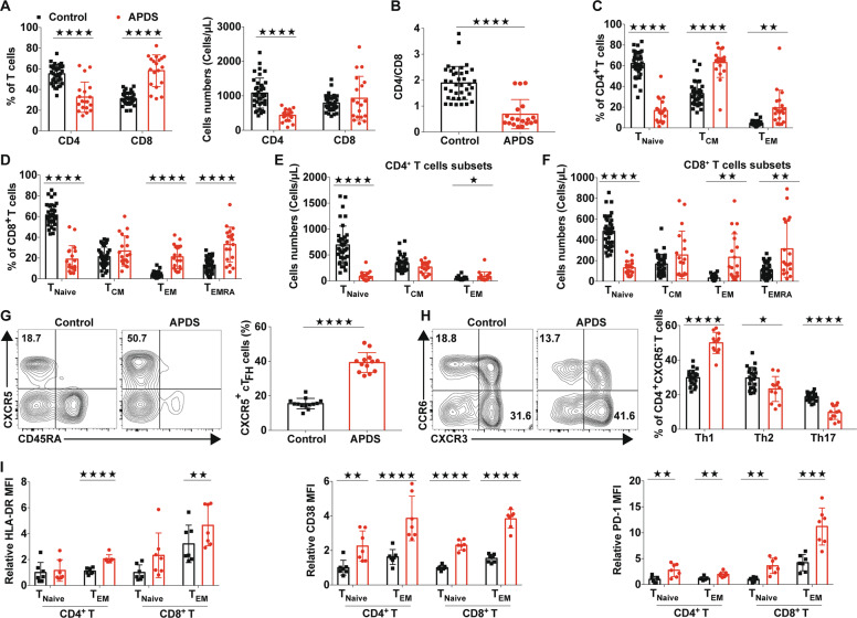
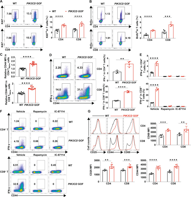
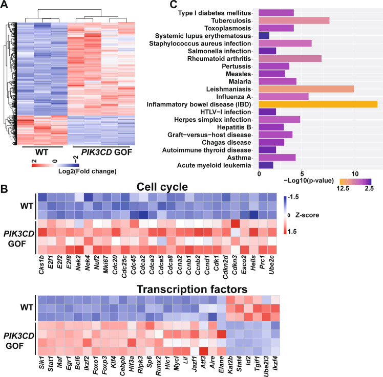
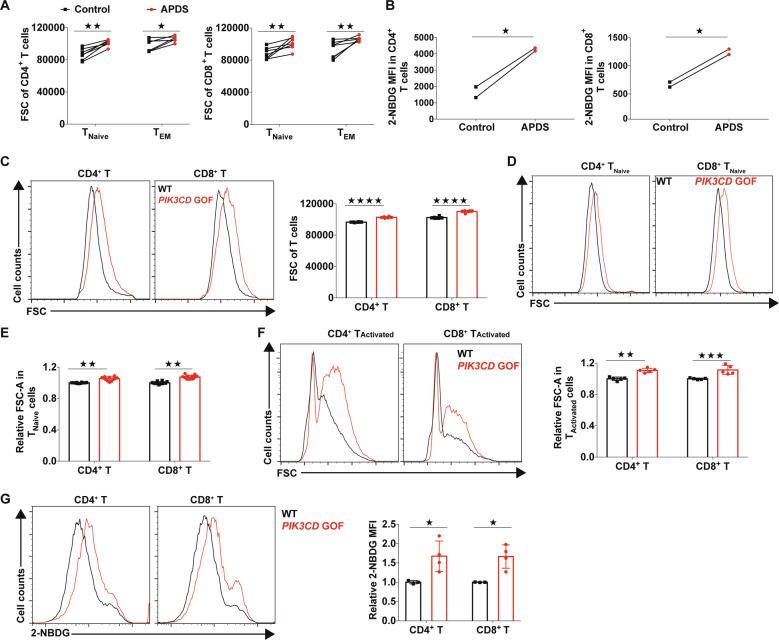
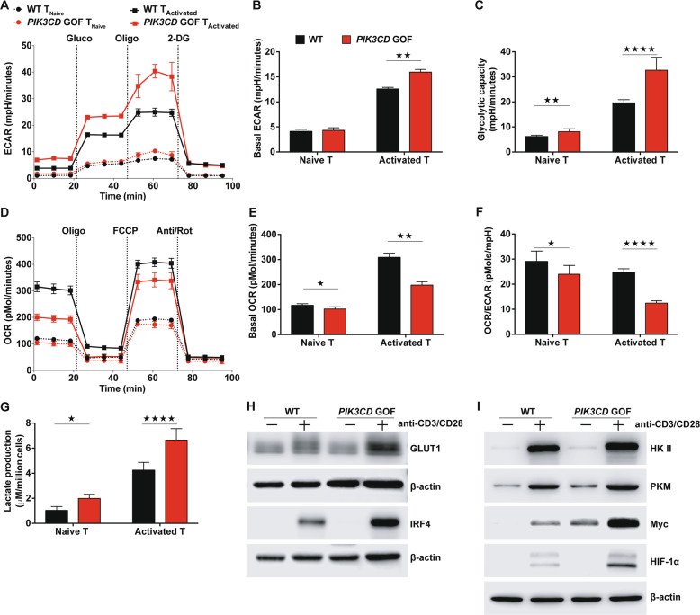
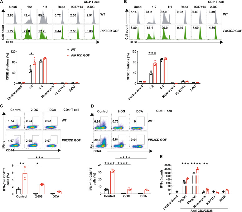
Activated phosphoinositide 3-kinase δ syndrome (APDS) is an autosomal-dominant combined immunodeficiency disorder resulting from pathogenic gain-of-function (GOF) mutations in the PIK3CD gene. Patients with APDS display abnormal T cell homeostasis. However, the mechanisms by which PIK3CD GOF contributes to this feature remain unknown. Here, with a cohort of children with PIK3CD GOF mutations from multiple regions of China and a corresponding CRISPR/Cas9 gene-edited mouse model, we reported that hyperactive PI3Kδ disrupted TNaive cell homeostasis in the periphery by intrinsically promoting the growth, proliferation, and activation of TNaive cells. Our results showed that PIK3CD GOF resulted in loss of the quiescence-associated gene expression profile in naive T cells and promoted naive T cells to overgrow, hyperproliferate and acquire an activated functional status. Naive PIK3CD GOF T cells exhibited an enhanced glycolytic capacity and reduced mitochondrial respiration in the resting or activated state. Blocking glycolysis abrogated the abnormal splenic T cell pool and reversed the overactivated phenotype induced by PIK3CD GOF in vivo and in vitro. These results suggest that enhanced aerobic glycolysis is required for PIK3CD GOF-induced overactivation of naive T cells and provide a potential therapeutic approach for targeting glycolysis to treat patients with APDS as well as other immune disorders.

Keywords: Activated phosphoinositide3-kinase δ syndrome; Aerobic glycolysis; Naive T cells; PIK3CD; Primary immunodeficiency disorders.


Conflict of interest statement

The authors declare no competing interests.

Figures

Fig. 1
Fig. 1
The T cell compartment in patients with APDS showed altered homeostasis. a The frequencies (left) and absolute numbers (right) of T cells in blood from patients with APDS (n = 19) and healthy controls (n = 38). (b) The ratio of CD4+ to CD8+ T cells in blood from patients with APDS (n = 19) and healthy controls (n = 38). c, d Proportions of naive T cell (TNaive; CD45RA+CCR7+), central memory T cell (TCM; CD45RACCR7+), effector memory T cell (TEM; CD45RACCR7), and CD45RA+ effector memory T cell (TEMRA; CD45RA+CCR7−) subsets in CD4+ and CD8+ T cell populations from patients with APDS (n = 19) and healthy controls (n = 38). e, f The absolute numbers of CD4+ and CD8+ T cell subsets from patients with APDS (n = 38) and healthy controls (n = 19). g Representative flow plots for the expression of CD45RA and CXCR5 within the CD3+ CD4+ T cells of the PBMCs and proportions of CD45RACXCR5+ cells (cTFH cells) within the CD3+CD4+ T cell populations from healthy controls and patients with APDS (n = 12). h Representative flow plots for the expression of CXCR3 and CCR6 (left) and the proportions of Th1 (CXCR3+CCR6), Th2 (CXCR3CCR6) and Th17 (CXCR3CCR6+) cells within the CD3+CD4+CXCR5CD45RA population (right) from healthy controls and untreated patients with APDS (n = 12 per group). i Expression of HLA-DR, CD38, PD-1 and CCR7 in CD4+ and CD8+ T cells from healthy controls (n = 7) and patients with APDS (n = 7). Numbers adjacent to outlined areas or in the indicated quadrants represent the percentage of cells in the area. Each symbol represents an individual throughout. Data are shown as the mean ± SD.P < 0.05, ★★P < 0.01, ★★★P < 0.001, and ★★★★P < 0.0001 determined by Students unpaired t-test
Fig. 2
Fig. 2
TNaive cells from PIK3CD GOF mice showed loss of quiescence and overactivation. a Flow cytometry analysis of the proportion of Ki67+ cells in splenic T cells from WT or PIK3CD GOF mice (n = 7 per group). b Flow cytometry analysis of the proportion of BrdU+ T cells in splenocytes from WT or PIK3CD GOF mice (n = 5 per group). c Expression of CD44 in naive T cells from WT or PIK3CD GOF mice (n = 12–13 per group). d Proportion of IFN-γ-producing T cells from WT or PIK3CD GOF mice. Isolated total T cells were stimulated by anti-CD3 and anti-CD28 antibodies for 24 h, and IFN-γ+ T cells were analyzed by flow cytometry (n = 4 per group). e, f Naive T cells from WT and PIK3CD GOF mice were stimulated with anti-CD3 and anti-CD28 antibodies for 24 h in the presence or absence of rapamycin, IC-87114 or DMSO as a control (n = 4 per group), and the proportion of IFN-γ-producing cells in CD4+ T cells (top) and CD8+ T cells (bottom) was determined by flow cytometry. g Expression of CD25, CD44 and CD69 in TCR-stimulated TNaive cells from WT or PIK3CD GOF mice (n = 5–8 per group). Numbers adjacent to outlined areas represent the percentage of cells in the area. Each symbol represents an individual throughout. Data are shown as the mean ± SD.P < 0.05, ★★P < 0.01, and ★★★★P < 0.0001 determined by Student’s unpaired t-test
Fig. 3
Fig. 3
The gene expression patterns in naive PIK3CD GOF T cells were altered. a Heatmap analysis of genes differentially expressed in CD4+ TNaive cells from WT and PIK3CD GOF mice (differentially expressed genes were identified with a false discovery rate ≤0.05 and fold change ≥2, n = 3–4 per group). b Genes differentially expressed were analyzed by gene ontology and grouped as cell cycle and transcription factors. c KEGG analysis of the most enriched pathways for differentially expressed genes in isolated CD4+ TNaive cells between WT and PIK3CD GOF mice
Fig. 4
Fig. 4
PIK3CD GOF led to increased growth and glucose uptake of T cells. a Size of isolated T cells from newly diagnosed patients with APDS and healthy controls. Cell size was evaluated by the forward-scatter (FSC) area. The patients shown here have not been treated with glucocorticoids or immunosuppressive agents (n = 7). b The glucose uptake of peripheral blood T cells was determined by staining with the glucose analog 2-NBDG. c–f Cell size of T cells from WT or PIK3CD GOF mice (n = 5–12 per group). Size of freshly isolated T cells (c), naive T cells (d, e), and T cells activated by anti-CD3/28 for 24 h (f). g Staining of 2-NBDG for assessing glucose uptake in naive T cells stimulated by anti-CD3 and anti-CD28 antibodies for 24 h (n = 3–4 per group). Each symbol represents an individual throughout. Data are shown as the mean ± SD. P < 0.05, ★★★P < 0.001 and ★★★★P < 0.001 determined by Student’s unpaired t-test
Fig. 5
Fig. 5
Peripheral T cells from PIK3CD GOF mice showed an elevated glycolysis capacity. ac The extracellular acidification rate (ECAR) was determined following consecutive injections of D-glucose (Gluco), the mitochondrial inhibitor oligomycin (Oligo) and the glycolytic inhibitor 2-DG. The time course of the ECAR (a), the basal ECAR (b) and calculations of glycolytic capacity (c) are shown. df The oxygen consumption rate (OCR) was determined following consecutive injections of the mitochondrial inhibitor oligomycin (Oligo), trifluoromethoxy carbonylcyanide phenylhydrazone (FCCP), and antimycin A and rotenone (Act/Rot). The time course of the OCR (d), the basal OCR (e) and the ratio of the OCR to the ECAR (f) are shown. g Lactate production in medium from naive T cells or activated T cells obtained from WT or PIK3CD GOF mice. h, i Immunoblot analysis of the levels of Glut1, IRF4, HKII, PKM, Myc and HIF-1α in naive T cells from WT or PIK3CD GOF mice left unstimulated or stimulated with anti-CD3 and anti-CD28 antibodies for 14 h. Data are shown as the mean ± SD for 3–4 independent experiments.P < 0.05, ★★P < 0.01, and ★★★★P < 0.0001 determined by Student’s unpaired t-test
Fig. 6
Fig. 6
Inhibiting glycolysis abolished T cell proliferation and activation caused by PIK3CD GOF. a, b CFSE-labeled TNaive cells from WT and PIK3CD GOF mice were stimulated with anti-CD3 and anti-CD28 antibodies for 72 h in the presence or absence of rapamycin, IC87114, 2-DG or vehicle as a control, and CFSE dilutions in (a) CD4+ T cells and (b) CD4+ T cells were determined by flow cytometry (n = 4 per group). c, d Naive T cells from WT and PIK3CD GOF mice were stimulated with anti-CD3 and anti-CD28 antibodies for 24 h in the presence or absence of 2-DG, DCA or vehicle as a control, and the proportion of IFN-γ-producing cells in c CD4+ T cells and d CD4+ T cells was determined by flow cytometry (n = 4 per group). e Naive T cells from WT and PIK3CD GOF mice were stimulated with anti-CD3 and anti-CD28 antibodies for 24 h in the presence of rapamycin, IC87114, 2-DG or vehicle as a control. After that, the concentration of IFN-γ in the medium was determined by ELISA (n = 5–6 per group). Solid black squares denote the concentration of IFN-γ in the medium below the detection limit. Numbers adjacent to outlined areas represent the percentage in the area. Data are shown as the mean ± SD for 3–6 independent experiments. P < 0.05, ★★P < 0.01, and ★★★★P < 0.0001 determined by two-way ANOVA
Fig. 7
Fig. 7
Inhibiting glycolysis with 2-DG reversed abnormal T cell homeostasis caused by PIK3CD GOF in vivo. a, b Proportions of TNaive and TEM cells within splenic CD4+ T cells (a) and CD8+ T cells (b). c, d Proportions of Ki67+ cells within splenic CD4+ T cells (c) and CD8+ T cells (d). e, f Proportions of CD69+ and CD25+ cells within splenic CD4+ T cells and CD8+ T cells. g, h Splenocytes from WT or PIK3CD GOF mice treated with vehicle or 2-DG were stimulated with PMA and ionomycin for 5 h, and the proportions of IFN-γ-producing T cells were analyzed by flow cytometry. Proportion of IFN-γ+ cells within CD4+ (g) and CD8+ T cells (i). Each symbol represents an individual mouse. Data are shown as the mean ±SD. ★★P < 0.01, ★★★P < 0.001 and, ★★★★P < 0.0001 determined by two-way ANOVA

Similar articles

Cited by

References

    1. Surh CD, Sprent J. Homeostasis of naive and memory T cells. Immunity. 2008;29:848–862. - PubMed
    1. Takada K, Jameson SC. Naive T cell homeostasis: from awareness of space to a sense of place. Nat. Rev. Immunol. 2009;9:823–832. - PubMed
    1. Silva SL, Sousa AE. Establishment and maintenance of the human naïve CD4+ T-cell compartment. Front Pediatr. 2016;4:1–10. - PMC - PubMed
    1. Angulo I, et al. Phosphoinositide 3-kinase δ gene mutation predisposes to respiratory infection and airway damage. Science. 2013;342:866–871. - PMC - PubMed
    1. Lucas CL, et al. Dominant-activating germline mutations in the gene encoding the PI(3)K catalytic subunit p110δ result in T cell senescence and human immunodeficiency. Nat. Immunol. 2014;15:88–97. - PMC - PubMed

Publication types

Substances