. 2020 Mar 23;11(3):199.
doi: 10.1038/s41419-020-2372-9.

Myofibroblast induces hepatocyte-to-ductal metaplasia via laminin-ɑvβ6 integrin in liver fibrosis

Affiliations

Myofibroblast induces hepatocyte-to-ductal metaplasia via laminin-ɑvβ6 integrin in liver fibrosis

Ting Xu et al. Cell Death Dis. .

Abstract

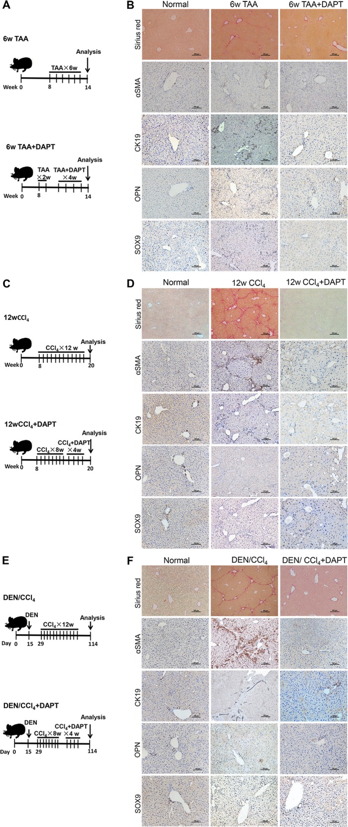
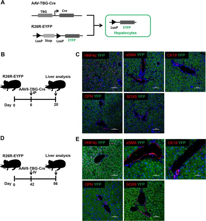
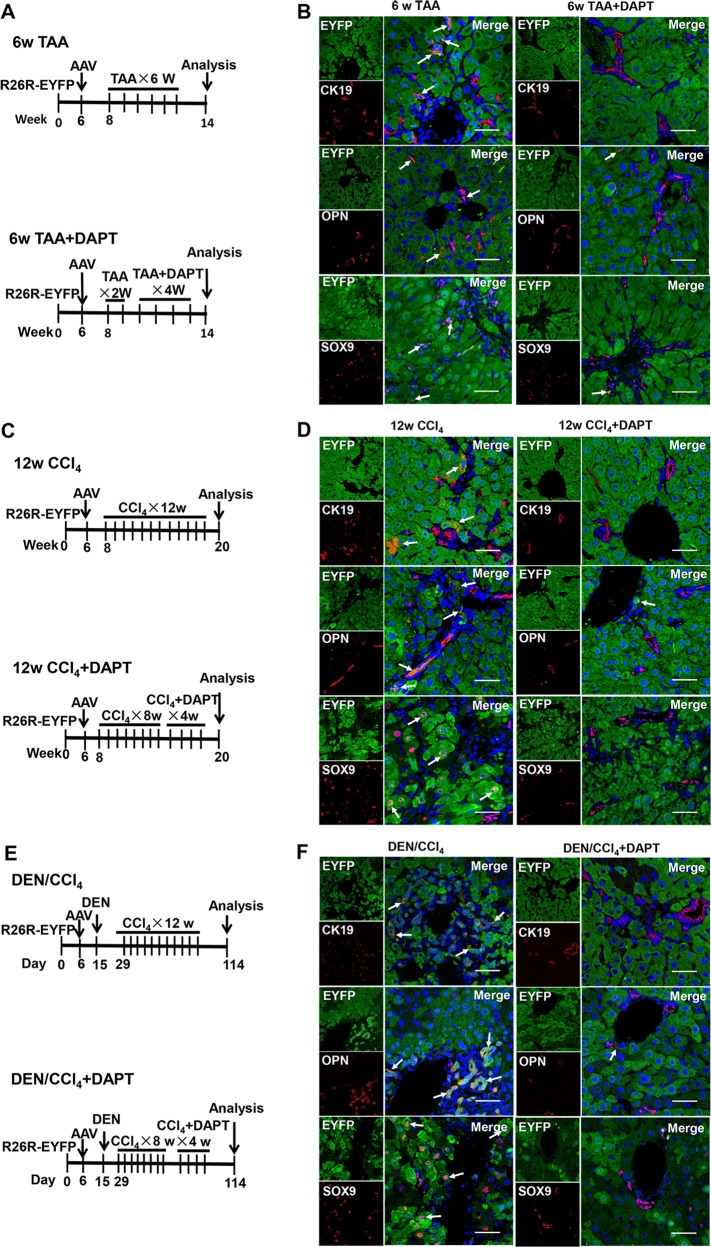
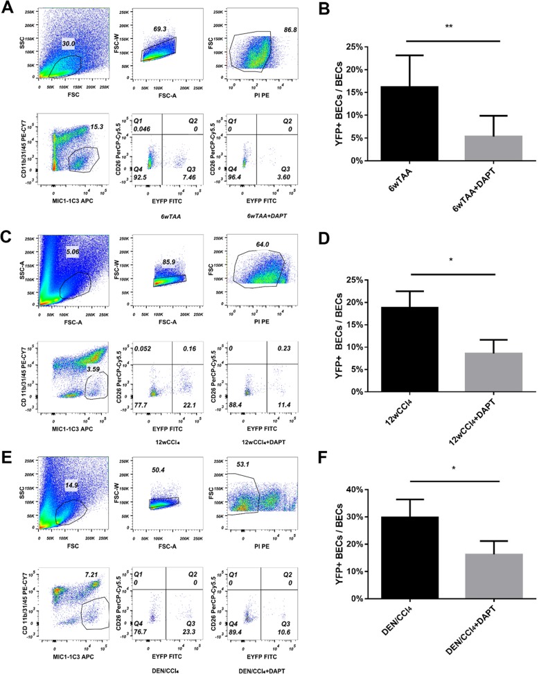
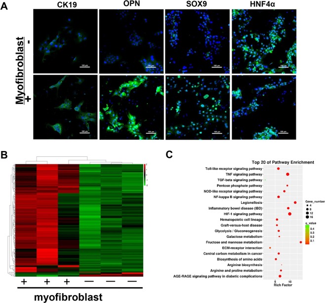
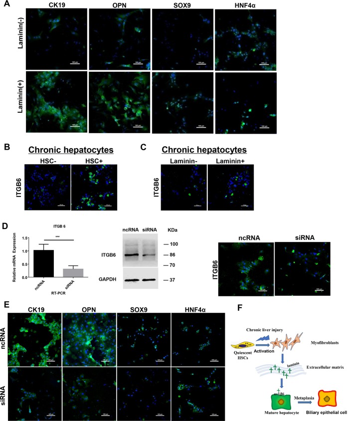
Hepatocytes undergo the metaplasia into ductal biliary epithelial cells (BECs) in response to chronic injury, and subsequently contribute to liver regeneration. The mechanism underlying hepatocyte-to-ductal metaplasia has not been explored until now. In mouse models of liver fibrosis, a florid BEC response was observed in fibrotic liver, and the depletion of myofibroblasts attenuated BEC expansion remarkably. Then, in hepatocyte fate-tracing mouse model, we demonstrated the conversion of mature hepatocytes into ductal BECs in fibrotic liver, and the depletion of myofibroblasts diminished the hepatocyte-to-ductal metaplasia. Finally, the mechanism underlying the metaplasia was investigated. Myofibroblasts secreted laminin-rich extracellular matrix, and then laminin induced hepatocyte-to-ductal metaplasia through ɑvβ6 integrin. Therefore, our results demonstrated myofibroblasts induce the conversion of mature hepatocytes into ductal BECs through laminin-ɑvβ6 integrin, which reveals that the strategy improve regeneration in fibrotic liver through the modification of specific microenvironment.


Conflict of interest statement

The authors declare that they have no conflict of interest.

Figures

Fig. 1
Fig. 1. Inhibition of HSC activation diminished the expansion of ductal biliary epithelial cells (BECs).
a Experiment design for TAA-induced liver fibrosis and DAPT-mediated inhibition of HSC activation in vivo. Vertical lines represent weekly intraperitoneal injections of TAA or TAA/DAPT. TAA thioacetamide, DAPT N-[N-(3,5-difluorophenacetyl)-l-alanyl]-S-phenylglycinet-butylester. b Representative liver sections from TAA-treated mice administrated with DAPT or the control (Sirius red staining, immunohistochemical staining). Collagen deposition was determined by Sirius red staining, immunohistochemical staining showed the expression of myofibroblasts (ɑSMA) and ductal BECs (CK19, OPN, and SOX9). c Experiment design for CCl4-induced liver fibrosis and DAPT-mediated inhibition of HSC activation in vivo. d Representative liver sections from CCl4-treated mice administrated with DAPT or the control (Sirius red staining, immunohistochemical staining). e Experiment design for DEN/CCl4-induced liver fibrosis and DAPT-mediated inhibition of HSC activation in vivo. DEN N-Nitrosodiethylamine. f Representative liver sections from DEN/CCl4-treated mice administrated with DAPT or the control (Sirius red staining, immunohistochemical staining).
Fig. 2
Fig. 2. Lineage-tracing of mature hepatocytes through the administration of AAV virus into the R26R-EYFP reporter mice.
a Schematic diagram of hepatocyte fate-tracing strategy through the injection of AAV8-TBG-Cre into R26R-EYFP (RosaYFP) mice. b Experiment design for lineage-tracing of mature hepatocytes in 6-day-old RosaYFP mice. 6-day-old RosaYFP mice were intraperitoneally injected with AAV8-TBG-Cre. c Immunofluorescence staining demonstrated AAV8-TBG-Cre efficiently labeled mature hepatocytes in 6-day-old RosaYFP mice. d Experiment design for lineage-tracing of mature hepatocytes in 6-week-old RosaYFP mice. RosaYFP mice (6-week-old) were intravenously injected with AAV8-TBG-Cre. e Immunofluorescence staining demonstrated AAV8-TBG-Cre efficiently labeled hepatocytes in 6-week-old RosaYFP mice.
Fig. 3
Fig. 3. Myofibroblasts induce the conversion of mature hepatocytes into ductal biliary epithelial cells in vivo.
a Experiment design for DAPT-mediated inhibition of HSC activation in TAA-treated RosaYFP reporter mice. b Immunofluorescent staining of fibrotic liver from TAA-treated RosaYFP reporter mice. Immunostaining of ductal BECs marker (CK19, OPN, and SOX9) in liver of RosaYFP mice showed that the percentage of hepatocytes-derived ductal BECs was diminished after DAPT-mediated inhibition of HSC activation in TAA-treated mice. c Experiment design for DAPT-mediated inhibition of HSC activation in CCl4-treated RosaYFP reporter mice. d Immunofluorescence staining of fibrotic liver from CCl4-treated RosaYFP reporter mice. e Experiment design for DAPT-mediated inhibition of HSC activation in DEN/CCl4-treated RosaYFP reporter mice. f Immunofluorescence staining of fibrotic liver from DEN/CCl4-treated RosaYFP reporter mice. Magnification, ×600.
Fig. 4
Fig. 4. Flow cytometry analysis demonstrated the inhibition of HSC activation diminished the conversion of mature hepatocytes into ductal BECs in vivo.
a Flow cytometry analysis showed hepatocyte-derived ductal BECs diminished in TAA-treated RosaYFP reporter mice upon DAPT-mediated inhibition of HSC activation. MIC1-1C3 was used as a surface marker of ductal BECs. b The percentage of ductal BECs derived from YFP-marked hepatocytes decreased significantly in TAA-treated RosaYFP reporter mice upon DAPT treatment. c Flow cytometry analysis showed hepatocyte-derived ductal BECs diminished in CCl4-treated RosaYFP reporter mice upon DAPT-mediate inhibition of HSC activation. d The percentage of ductal BECs derived from YFP-marked hepatocytes decreased significantly in CCl4-treated RosaYFP reporter mice upon DAPT treatment. e Flow cytometry analysis showed hepatocyte-derived ductal BECs diminished in DEN/CCl4-treated RosaYFP reporter mice upon DAPT-mediate inhibition of HSC activation. f The percentage of ductal BECs derived from YFP-marked hepatocytes decreased significantly in DEN/CCl4-treated RosaYFP reporter mice after the treatment of DAPT. Each bar represents the mean ± SD for at least triplicate experiments and the P-value was determined by Student’s t-test (***P < 0.001, **P < 0.01, *P < 0.05).
Fig. 5
Fig. 5. Myofibroblasts induce the conversion of mature hepatocytes into ductal biliary epithelial cells in vitro through co-culture of myofibroblasts and chronic injured hepatocytes.
Immunofluorescence staining and RNA-sequencing were performed to determine the effect of myofibroblasts on biological characteristic of chronic injured hepatocytes. a Immunofluorescence staining showed the expression of ductal BECs marker increased in chronic injured hepatocytes upon the treatment of myofibroblasts. b Heat map of the differentially expressed mRNAs in chronic injured hepatocytes treated with myofibroblasts. c Pathway enrichment analysis was performed to identify pathways involved in the conversion of mature hepatocytes into ductal biliary epithelial cells.
Fig. 6
Fig. 6. Myofibroblasts induce the conversion of mature hepatocytes into ductal biliary epithelial cells through the interaction of laminin–β6 integrin.
a Up-regulation of ductal BECs marker (CK19, OPN, and SOX9) and down-regulation of hepatocytes marker (HNF4ɑ) were observed in chronic injured hepatocytes upon the treatment of laminin. b Up-regulation of integrin ɑvβ6 expression in chronic injured hepatocytes upon the treatment of myofibroblasts. c Up-regulation of integrin ɑvβ6 expression in chronic injured hepatocytes upon the treatment of laminin. d siRNA-mediated inhibition of integrin ɑvβ6 expression in chronic injured hepatocytes. Primary hepatocytes isolated from fibrotic liver were transfected with 50 nM of ITGB6-siRNA and control scrambled siRNA (NC), qRT-PCR, western Blot and IF staining demonstrated efficient knockdown of ITGB6 expression through siRNA. e Immunofluorescence staining showed down-regulation of ductal BECs markers (CK19, OPN, and SOX9) expression and up-regulation of HNF4ɑ expression in primary hepatocytes transfected with ITGB6-siRNA compared with the control. f Graphical summary of signaling pathway in which myofibroblasts induce the conversion of mature hepatocytes into LPCs through the interaction of laminin–integrin ɑvβ6. Each bar represents the mean ± SD for at least triplicate experiments and the P-value was determined by Student’s t-test (***P < 0.001, **P < 0.01, *P < 0.05).

Similar articles

Cited by

References

    1. Diehl AM, Chute J. Underlying potential: cellular and molecular determinants of adult liver repair. J. Clin. Investig. 2013;123:1858–1860. doi: 10.1172/JCI69966. - DOI - PMC - PubMed
    1. Itoh T, Miyajima A. Liver regeneration by stem/progenitor cells. Hepatology. 2014;59:1617–1626. doi: 10.1002/hep.26753. - DOI - PubMed
    1. Huch M. Regenerative biology: the versatile and plastic liver. Nature. 2015;517:155–156. doi: 10.1038/517155a. - DOI - PubMed
    1. Shang H, Wang Z, Song Y. Liver progenitor cells-mediated liver regeneration in liver cirrhosis. Hepatol. Int. 2016;10:440–447. doi: 10.1007/s12072-015-9693-2. - DOI - PubMed
    1. Hindley CJ, Mastrogiovanni G, Huch M. The plastic liver: differentiated cells, stem cells, every cell? J. Clin. Investig. 2014;124:5099–5102. doi: 10.1172/JCI78372. - DOI - PMC - PubMed

Publication types