. 2020 Jul 1;130(7):3833-3847.
doi: 10.1172/JCI123353.

Circular RNA-ZNF532 regulates diabetes-induced retinal pericyte degeneration and vascular dysfunction

Affiliations

Circular RNA-ZNF532 regulates diabetes-induced retinal pericyte degeneration and vascular dysfunction

Qin Jiang et al. J Clin Invest. .

Abstract

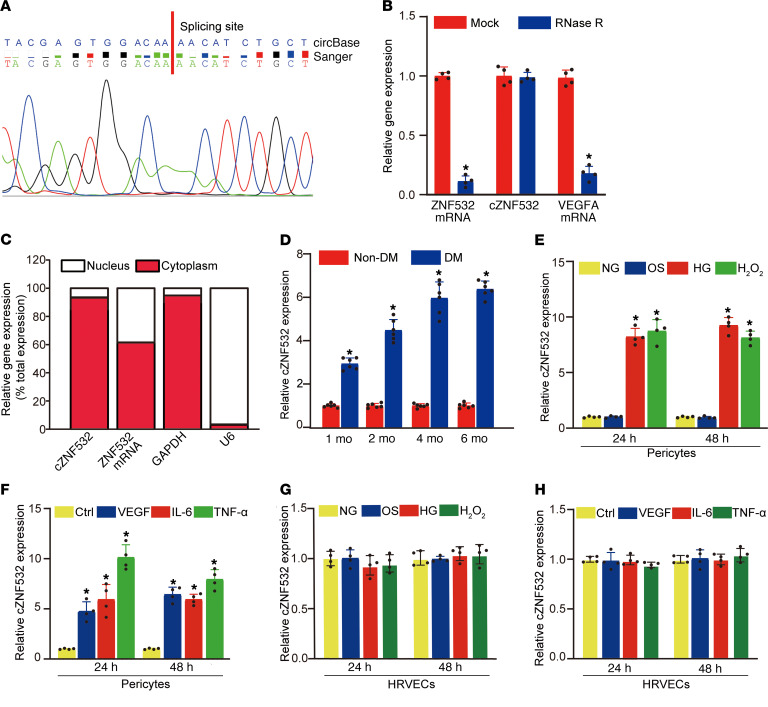
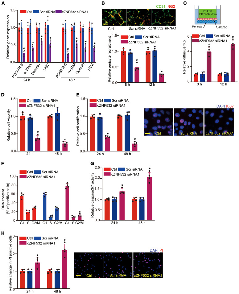
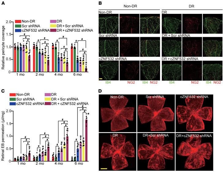
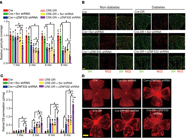
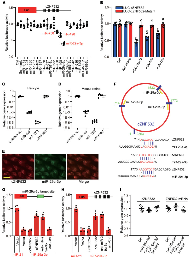
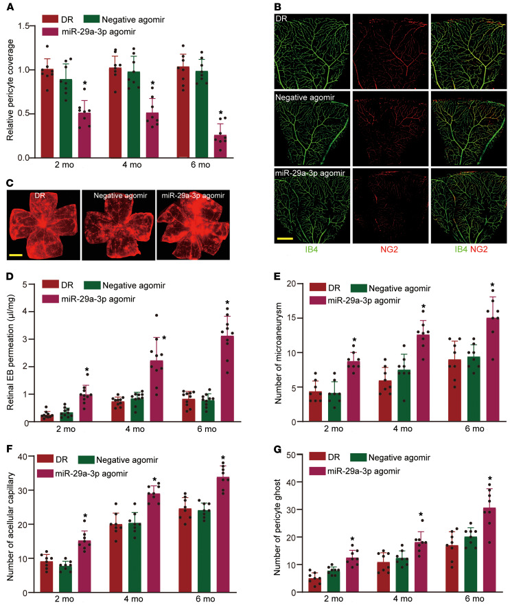
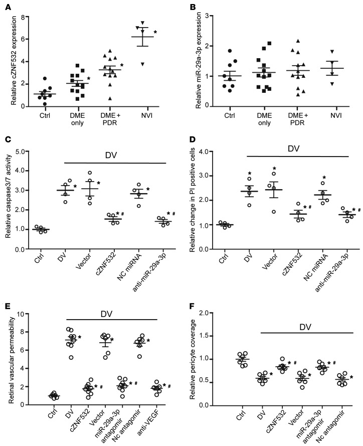
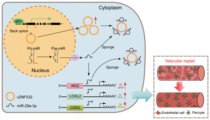
Diabetic retinopathy (DR) is the leading cause of blindness in working-age adults. Vascular pericyte degeneration is the predominant clinical manifestation of DR, yet the mechanism governing pericyte degeneration is poorly understood. Circular RNAs (circRNAs) play important roles in multiple biological processes and disease progression. Here, we investigated the role of circRNA in pericyte biology and diabetes-induced retinal vascular dysfunction. cZNF532 expression was upregulated in pericytes under diabetic stress, in the retinal vessels of a diabetic murine model, and in the vitreous humor of diabetic patients. cZNF532 silencing reduced the viability, proliferation, and differentiation of pericytes and suppressed the recruitment of pericytes toward endothelial cells in vitro. cZNF532 regulated pericyte biology by acting as a miR-29a-3p sponge and inducing increased expression of NG2, LOXL2, and CDK2. Knockdown of cZNF532 or overexpression of miR-29a-3p aggravated streptozotocin-induced retinal pericyte degeneration and vascular dysfunction. By contrast, overexpression of cZNF532 or inhibition of miR-29a-3p ameliorated human diabetic vitreous-induced retinal pericyte degeneration and vascular dysfunction. Collectively, these data identify a circRNA-mediated mechanism that coordinates pericyte biology and vascular homeostasis in DR. Induction of cZNF532 or antagonism of miR-29a-3p is an exploitable therapeutic approach for the treatment of DR.

Keywords: Diabetes; Noncoding RNAs; Ophthalmology; Retinopathy.


Conflict of interest statement

Conflict of interest: The authors have declared that no conflict of interest exists.

Figures

Figure 1
Figure 1. Identification of cZNF532 as a high glucose–regulated circRNA in pericytes.
(A) Sanger sequencing was conducted to detect cZNF532 expression in pericytes. The result of Sanger sequencing (bottom) was consistent with the sequence of cZNF532 in circBase (top). (B) Total RNAs were digested with RNAse R followed by qRT-PCR detection of cZNF532 expression. ZNF532 mRNA and VEGFA mRNA was detected as the RNAse R–sensitive control (n = 4, Student’s t test). (C) The expression of nucleus transcript (U6), cytoplasm transcript (GAPDH), ZNF532 mRNA, and cZNF532 was detected by qRT-PCRs in the nucleus and cytoplasm fraction of pericytes (n = 4). (D) cZNF532 expression was detected by qRT-PCRs in retinal vessels isolated from nondiabetic retinas and diabetic retinas 1 month, 2 months, 4 months, and 6 months after the induction of diabetes (n = 6 animals per group, Student’s t test). The blood glucose levels of diabetic mice were above 300 mg/dL. (E and G) cZNF532 expression was detected by qRT-PCRs in pericytes (E) or HRVECs (G) cultured in medium containing normal glucose (NG, 5.55 mM), 5.55 mM glucose plus 24.45 mM pyruvate (osmotic control, OS), high glucose (HG, 30 mM), or H2O2 (100 μm) for 24 hours and 48 hours. (F and H) cZNF532 expression was detected by qRT-PCRs in pericytes (F) or HRVECs (H) cultured in the medium containing VEGF (20 ng/mL), IL-6 (50 ng/mL), and TNF-α (50 ng/mL) or left untreated (Ctrl) for 24 hours and 48 hours. For EH, *P < 0.05, 1-way ANOVA followed by Bonferroni’s post hoc comparison test, error bar indicates SD.
Figure 2
Figure 2. cZNF532 regulates pericyte function in vitro.
(A) The expression of pericyte markers, including PDGFR-β, α-SMA, desmin, and NG2, was detected by qRT-PCRs in pericytes after the transfection of scrambled (Scr) siRNA or cZNF532 siRNA1, or left untreated (Ctrl) for 24 hours and 48 hours (n = 4). (B) WT (Ctrl), cZNF532 siRNA1, or Scr siRNA–transfected pericytes were cocultured with HRVECs for 8 hours or 12 hours and then stained with NG2 (pericytes) and CD31 (HRVECs) to detect the recruitment of pericytes toward HRVECs (n = 4). The representative images at 12 hours are shown. Scale bar: 100 μm. (C) WT (Ctrl), cZNF532 siRNA1, or Scr siRNA–transfected pericytes were cocultured with HRVECs for 8 hours and 12 hours. Fluorescent solute (FITC-Dextran, 70 kDa) was added to the apical chamber and the rate of flux across the HRVEC monolayer was detected by a microplate reader. Endothelial barrier permeability was shown as relative diffusive flux change (n = 4). Average Po for the control samples at 8 hours and 12 hours was 2.78 × 10⁻⁶ cm/sec and 1.62 × 10⁻⁶ cm/sec, respectively. (D and E) Pericytes were transfected with Scr siRNA or cZNF532 siRNA1, or left untreated (Ctrl) for 24 hours or 48 hours. Cell viability was detected by MTT method (D, n = 4). Cell proliferation was detected by Ki67 staining. The representative images at 48 hours were shown (E, scale bar: 20 μm, n = 4). (F) Cell cycles of pericytes were detected by flow cytometry 48 hours after the transfection of Scr siRNA or cZNF532 siRNA1. Cell percentage in each phase was calculated by BD Cell Quest Pro software. (G and H) Pericytes were transfected with Scr siRNA or cZNF532 siRNA1, or left untreated (Ctrl), and then exposed to 30 mM glucose for 24 hours or 48 hours. Apoptotic cells were detected by caspase-3/7 activity (G, n = 4) or PI staining (H, n = 4). Representative PI staining images at 48 hours were shown (scale bar: 100 μm). All significant difference was determined by 1-way ANOVA followed by Bonferroni’s post hoc test. Error bar indicates SD. *P < 0.05.
Figure 3
Figure 3. cZNF532 regulates retinal pericyte function and vascular integrity in vivo.
(A and B) Pericyte coverage was quantified by staining the whole-mount retina with Isolectin IB4 and NG2 in the nondiabetic C57BL/6 mice (non-DR) or diabetic mice (Cre-DR) without or with intravitreous injection of Scr shRNA or cZNF532 shRNA after 1 month, 2 months, 4 months, or 6 months of treatment. To visualize a whole leaf of retinal vessel, tile scanning was used whereby multiple overlapping (10%–20% overlap) images were captured by a ×10 lens with identical gain setting. The composite images were generated by arraying the individual images in Adobe Photoshop. The statistical result (A, n = 8) and representative composite images after 6 months of treatment are shown (B, scale bar: 100 μm). (C and D) The mice were infused with Evans blue (EB) dye for 2 hours. The tile-scanning images of whole retinal vessels were taken using a ×4 lens with identical gain settings. The statistical result of EB extravasation (C, n = 8) and representative images of flat-mounted retinas after 6 months of treatment (D, scale bar: 500 μm). The red fluorescence indicates EB signal. The blood glucose levels of diabetic mice were above 300 mg/dL. All significant differences were evaluated by Mann-Whitney U test or Kruskal-Wallis’s test followed by Bonferroni’s post hoc test. Error bar indicates SD. *P < 0.05 compared with non-DR group; #P < 0.05 between the marked groups.
Figure 4
Figure 4. Conditional knockdown of cZNF532 in pericytes induces retinal vascular dysfunction in vivo.
(A and B) Pericyte coverage was quantified by staining the whole-mount retinas with Isolectin IB4 and NG2 in nondiabetic C57BL/6 Cre mice (Cre) or diabetic Cre mice (Cre-DR) without or with intravitreous injection of Scr shRNA or cZNF532 shRNA after 1, 2, 4, or 6 months of treatment (n = 8; scale bar: 100 μm). Tile scanning and Adobe Photoshop were used to generate the composite images of a whole leaf of retinal vessels. The statistical result and representative images after 6 months of treatment are shown. (C and D) The mice were infused with EB dye for 2 hours. The tile-scanning images of whole retinal vessels were taken using a ×4 lens with identical gain settings. The statistical result of EB extravasation and representative images of flat-mounted retinas after 6 months of treatment (n = 8; scale bar, 500 μm). The red fluorescence indicates EB signal. The blood glucose levels of diabetic mice were above 300 mg/dL. All significant differences were evaluated by Mann-Whitney U test or Kruskal-Wallis test followed by Bonferroni’s post hoc test. Error bar indicates SD. *P < 0.05 compared with Cre group. #P < 0.05 among the marked groups.
Figure 5
Figure 5. cZNF532 regulates pericyte function by acting as a miRNA sponge in vitro.
(A) HEK293T cells were transfected with pGL3-Basic (Ctrl) or LUC-cZNF532 with different miRNA mimic and pRL-TK vector. pRL-TK vector was transfected as the internal transfection control. Luciferase assays were conducted 48 hours after transfection using the Dual-Luciferase Reporter Assay kit. Normalized value of luciferase activity for Ctrl group was set to 1 (n = 4). (B) LUC-cZNF532 or LUC-cZNF532-mutant was cotransfected without or with miRNA mimic and pRL-TK vector. Luciferase assays were conducted 48 hours after transfection. Normalized value of luciferase activity was set to 1 in cells transfected with LUC-cZNF532 and pRL-TK (n = 4). (C and D) Relative expression abundance of cZNF532, miR-29a-3p, miR-498, and miR-758 was detected by qRT-PCR in pericytes (C, n = 4) and mouse retinas (D, n = 6). (E) Expression distribution of cZNF532 and miR-29a-3p in pericytes was detected by RNA-FISH assay (scale bar: 20 μm). (F) The schematic figure shows the putative binding sites of miR-29a-3p on cZNF532 transcript. (G and H) Luciferase reporter with perfect miR-29a-3p target site (G) or entire cZNF532 sequence was constructed (H). The constructed reporter was transfected with 40 ng empty vector (vector, pcDNA3), 40 ng cZNF532-ir (pcDNA3-cZNF532-ir), 40 ng cZNF532 (pcDNA3-cZNF532), 5 nM anti–miR-29a-3p (anti–miR-29a-3p), or 5 nM anti–miRNA control (anti-Ctl), together with 20 ng pJEBB-miR-29a-3p or pJEBB-miR-21 expression plasmid. Meanwhile, pRL-TK vector was transfected as the internal control. The normalized value of luciferase activity was set to 1 in cells transfected with the constructed Luc reporter, pcDNA3 (vector), pJEBB-miR-21, and pRL-TK. Luciferase activity was detected at 48 hours after transfection (n = 4, *P < 0.05 vs. miR-29a-3p plus vector). (I) Pericytes were transfected with miR-29a-3p mimic or miR-29a-3p inhibitor, or left untreated (Ctrl) for 48 hours. The expression of cZNF532 and ZNF532 mRNA was detected by qRT-PCR (n = 4). The significant difference was determined by 1-way ANOVA followed by Bonferroni’s post hoc test. Error bar indicates SD. *P < 0.05.
Figure 6
Figure 6. cZNF532-miR-29a-3p-NG2/LOXL2/CDK2 signaling regulates retinal pericyte function and vascular integrity in vivo.
(A and B) Diabetic C57BL/6 mice (3 months old, male) received an intravitreous injection of miR-29a-3p agomir or negative control (NC) agomir, or were left untreated (Ctrl). Pericyte coverage was quantified by staining the whole-mount retinas with Isolectin IB4 and NG2 after 2 months, 4 months, and 6 months of treatment (n = 8; scale bar: 100 μm). The statistical result and representative images after 6 months of treatment are shown. (C and D) The above-mentioned mice were infused with EB dye for 2 hours. The tile-scanning images of entire retinal vessels were taken using a ×4 lens with identical gain settings. The representative images of flat-mounted retinas after 6 months of treatment (C, n = 10; scale bar: 500 μm) and statistical result of EB extravasation are shown (D). The red fluorescence indicates EB signal. (EG) Retinal trypsin digestion and PAS staining were conducted to detect the number of microaneurysms (E, n = 8 per mm2 retina), acellular capillaries (F, n = 8 per mm2 retina), and pericyte ghosts (G, n = 8 per mm2 retina). The blood glucose levels of diabetic mice were above 300 mg/dL. The significant difference was evaluated by Kruskal-Wallis’s test followed by Bonferroni’s post hoc test. Error bar indicates SD. *P < 0.05.
Figure 7
Figure 7. Clinical implication of cZNF532/miR-29a-3p/NG2, LOXL2, and CDK2 signaling in retinal vascular dysfunction.
(A and B) Human vitreous specimens were obtained from 36 subjects at the time of pars plana vitrectomy, including non-DR samples (Ctrl, n = 8 eyes) and those from patients with DME only (n = 12 eyes), DME with PDR (n = 12 eyes), and NVI (n = 4 eyes). qRT-PCR was conducted to detect the expression of cZNF532 (A) and miR-29a-3p (B) in vitreous samples. *P < 0.05 versus Ctrl group, Kruskal-Wallis’s test followed by Bonferroni’s post hoc test. (C and D) Pericytes were transfected with cZNF532 overexpression vector (cZNF532), null vector (Vector), anti–miR-29a-3p, or negative control miRNA for 24 hours or left untreated, and then incubated without (Ctrl) or with 50 μL diabetic vitreous (DV) for 24 hours. Cell apoptosis was detected by caspase-3/7 activity (C, n = 4) or PI staining (D, n = 4). *P < 0.05 versus Ctrl group, #P < 0.05 versus DV group. One-way ANOVA followed by Bonferroni’s post hoc test. (E) EB evaluation of retinal vasopermeability in retinal extracts from mice injected with PBS (Ctrl, n = 6), diabetic vitreous without (DV, n = 8) or with cZNF532 overexpression vector (cZNF532, n = 8), null vector (Vector, n = 6), miR-29a-3p antagomir (n = 8), negative control (NC, n = 6) antagomir, or anti-VEGF (n = 6) after 1 week of treatment. (F) Pericyte coverage was quantified by staining the whole-mount retinas with IB4 and NG2 from the mice after intravitreal injection of PBS (Ctrl, n = 6) diabetic vitreous without (DV, n = 6) or with cZNF532 overexpression vector (cZNF532, n = 6), null vector (Vector, n = 6), miR-29a-3p antagomir (n = 6), or NC (n = 6) antagomir 1-week after treatment. The statistical result was shown. *P < 0.05 versus Ctrl group, #P < 0.05 versus DV group, Kruskal-Wallis’s test followed by Bonferroni’s post hoc test. Error bar indicates SD.
Figure 8
Figure 8. Model of cZNF532 function and mechanism in retinal vascular dysfunction.
cZNF532, an important regulator of pericyte function and vascular homeostasis, protects against diabetes-induced retinal pericyte degeneration and vascular dysfunction by acting as a miR-29a-3p sponge to sequester and inhibit miR-29a-3p activity, inducing increased expression of NG2, LOXL2, and CDK2.

Similar articles

Cited by

References

    1. Geevarghese A, Herman IM. Pericyte-endothelial crosstalk: implications and opportunities for advanced cellular therapies. Transl Res. 2014;163(4):296–306. doi: 10.1016/j.trsl.2014.01.011. - DOI - PMC - PubMed
    1. Armulik A, Abramsson A, Betsholtz C. Endothelial/pericyte interactions. Circ Res. 2005;97(6):512–523. doi: 10.1161/01.RES.0000182903.16652.d7. - DOI - PubMed
    1. Bischoff FC, et al. Identification and functional characterization of hypoxia-induced endoplasmic reticulum stress regulating lncRNA (HypERlnc) in Pericytes. Circ Res. 2017;121(4):368–375. doi: 10.1161/CIRCRESAHA.116.310531. - DOI - PubMed
    1. Stark K, et al. Capillary and arteriolar pericytes attract innate leukocytes exiting through venules and ‘instruct’ them with pattern-recognition and motility programs. Nat Immunol. 2013;14(1):41–51. doi: 10.1038/ni.2477. - DOI - PubMed
    1. Hall CN, et al. Capillary pericytes regulate cerebral blood flow in health and disease. Nature. 2014;508(7494):55–60. doi: 10.1038/nature13165. - DOI - PMC - PubMed

Publication types