. 2020 May 21;11(1):2538.
doi: 10.1038/s41467-020-15935-0.

Raptor determines β-cell identity and plasticity independent of hyperglycemia in mice

Affiliations

Raptor determines β-cell identity and plasticity independent of hyperglycemia in mice

Qinglei Yin et al. Nat Commun. .

Abstract

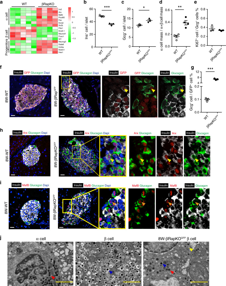
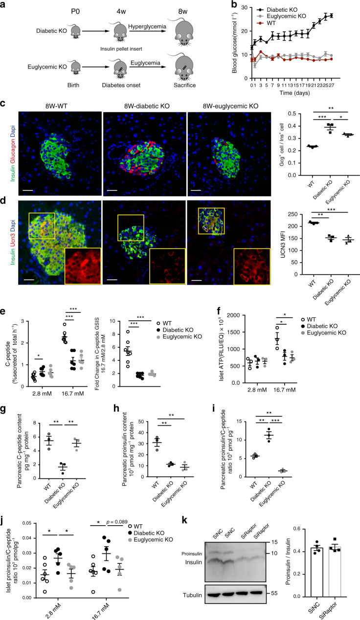
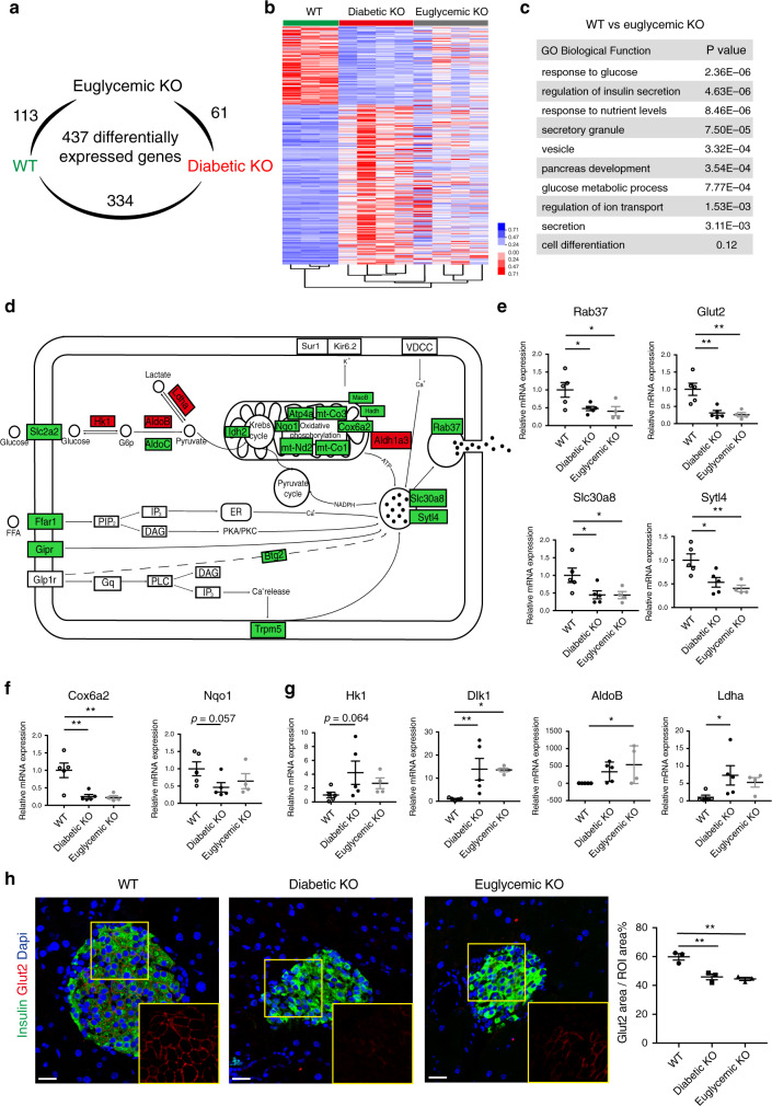
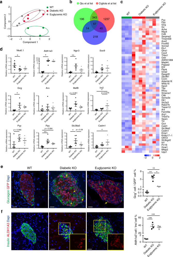
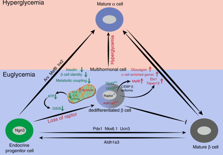
Compromised β-cell identity is emerging as an important contributor to β-cell failure in diabetes; however, the precise mechanism independent of hyperglycemia is under investigation. We have previously reported that mTORC1/Raptor regulates functional maturation in β-cells. In the present study, we find that diabetic β-cell specific Raptor-deficient mice (βRapKOGFP) show reduced β-cell mass, loss of β-cell identity and acquisition of α-cell features; which are not reversible upon glucose normalization. Deletion of Raptor directly impairs β-cell identity, mitochondrial metabolic coupling and protein synthetic activity, leading to β-cell failure. Moreover, loss of Raptor activates α-cell transcription factor MafB (via modulating C/EBPβ isoform ratio) and several α-cell enriched genes i.e. Etv1 and Tspan12, thus initiates β- to α-cell reprograming. The present findings highlight mTORC1 as a metabolic rheostat for stabilizing β-cell identity and repressing α-cell program at normoglycemic level, which might present therapeutic opportunities for treatment of diabetes.


Conflict of interest statement

The authors declare no competing interests.

Figures

Fig. 1
Fig. 1. Increased α/β cell ratio in βRapKOGFP mice.
a Heatmap of genes critical to endocrine cell in wild type (WT) and βRapKO islets (n = 4–5). b The number of Ins+ cells per islet (n = 3, p = 0.0008) and c the number of Gcg+ cells per islet were calculated (n = 3, p = 0.023). At least, 46 islets were used for quantifications. d The proportion of pancreatic α-cell mass among pancreatic α- and β-cell mass was shown (n = 4, p = 0.0017). e The proliferation of Gcg+ cell was determined by quantification of the percentage of Ki67+ cells in Gcg+ cells (n = 3). At least, 50 islets were used for quantifications. f Pancreatic sections from 8-week-old WT, βRapKOGFP mice were immunostained for insulin (white), glucagon (green), and GFP (red). Nuclei were stained with DAPI (blue) (n = 3). The yellow arrows indicated insulin, glucagon, and GFP co-stained cells. Scale bars, 20 μm. g The percentage of GFP+Gcg+ cells among GFP+ cells in 8-week-old WT and βRapKOGFP mice (n = 3, p = 1.83046E-05). At least, 1837 GFP+ cells over three mice each group were used for quantifications. h Representative pancreatic sections showed expression of α-cells marker—Arx (red), insulin (white), and glucagon (green) in WT and βRapKOGFP mice at 8 weeks of age (n = 3). Scale bars, 20 μm. i Immunostaining showed expression of α-cells marker—MafB (red), insulin (white), and glucagon (green) in control and βRapKOGFP mice at 8 weeks of age (n = 3). Yellow boxes showed the specific areas of the islet, which were enlarged and represented by arrows on the right to demonstrate protein expression within specific cells. Scale bars, 20 μm. j Transmission electron microscopy (TEM) of pancreatic islets reveals a combination of mature insulin, immature insulin, and glucagon-like granules in the same cell in βRapKOGFP mice (n = 3 independent samples). Granule identity is indicated by colored arrows: glucagon (red), mature insulin (blue), and immature insulin (yellow). Data represent means ± SEM. *p < 0.05, **p < 0.01, ***p < 0.001 by two-sided Student’s t-test.
Fig. 2
Fig. 2. Impaired proinsulin synthesis and ATP levels for GSIS.
a Schematic of insulin pump implantation. b Random blood glucose levels were monitored every other day in WT, diabetic βRapKOGFP, and euglycemic βRapKOGFP mice from 4 weeks to 8 weeks old (n = 6 for WT, n = 7 for diabetic KO, n = 13 for euglycemic KO). c Representative pancreatic sections immunostained for insulin (green) and glucagon (red). The ratio of Gcg+ cells to Ins+ cells was calculated (n = 3). At least 50 islets or 2000 insulin-positive cells were used for quantifications. Scale bars, 20 μm. d Images of islets labeled with insulin (green) and UCN3 (red). MFI of UCN3 in these three groups (n = 3). At least 10 islets from three sections were used for MFI. Yellow boxes showed the expression of UCN3. Scale bars, 20 μm. e In vitro glucose-stimulated C-peptide secretion in islets at 2.8 and 16.7 mM glucose levels for 1 h (reported as percent of C-peptide content) (n = 7 for WT, n = 6 for diabetic KO, n = 6 for euglycemic KO). White, black, and gray circles represented the WT, diabetic βRapKOGFP, and euglycemic βRapKOGFP groups, respectively. Stimulation index of C-peptide secretion in diabetic βRapKOGFP and euglycemic βRapKOGFP mice as compared with controls (n = 7 for WT, n = 6 for diabetic KO, n = 6 for euglycemic KO). f ATP content at 2.8 mM glucose and 16.7 mM glucose (n = 3). g Pancreatic C-peptide content (n = 3 independent samples) and h pancreatic proinsulin content normalized by protein concentration were shown (n = 3 independent samples). i The ratio of pancreatic proinsulin to C-peptide content was determined (n = 3 independent samples). j The ratio of islet proinsulin to C-peptide content per 10 size-matched islets at 2.8 mM and high 16.7 mM glucose levels was determined (n = 6 for WT, n = 5 for diabetic KO, n = 5 for euglycemic KO). k INS-1 cells were transfected with SiRaptor or SiNC for 72 h. Immunoblotting and quantification of insulin and proinsulin in INS-1 cells transfected with SiNC or SiRaptor (n = 4 independent cell experiments). Data represent means ± SEM. *p < 0.05, **p < 0.01, ***p < 0.001 by two-sided Student’s t-test and one-way ANOVA adjusted by LSD multiple comparison, p values included in source data.
Fig. 3
Fig. 3. Impaired metabolic coupling of insulin secretion.
a The number of differentially expressed genes within the three groups (n = 3–4). b Gene expression profiles regulated by Raptor were subjected to hierarchical clustering (n = 3–4). c GO analysis of differentially expressed genes as identified by RNA-seq of 8-week-old WT (n = 3) and euglycemic βRapKOGFP β-cells (n = 4) was associated with β-cell function. d Visualization of differential expression of genes involved in insulin secretion. e–g Relative expression of selected transcripts associated with β-cell secretion function (n = 5 independent sample for WT, n = 5 independent sample for diabetic βRapKOGFP, n = 4 independent sample for euglycemic βRapKOGFP, p values included in source data) (e), mitochondrial metabolism (n = 5 independent sample for WT, n = 5 independent sample for diabetic βRapKOGFP, n = 4 independent sample for euglycemic βRapKOGFP, p values included in source data) (f), and disallowed genes (n = 5 independent sample for WT, n = 5 independent sample for diabetic βRapKOGFP, n = 4 independent sample for euglycemic βRapKOGFP, p values included in source data) (g) in WT, diabetic βRapKOGFP, and euglycemic βRapKOGFP β-cells by qRT-PCR. h Representative immunofluorescent staining for Glut2 (red) and insulin (green) among 8-week-old WT, diabetic βRapKOGFP, and euglycemic βRapKOGFP mice (n = 3). Insets showed different expression levels of Glut2. Quantification of Glut2 areas in Ins+ areas in WT, diabetic βRapKOGFP, and euglycemic βRapKOGFP mice (n = 3, WT vs diabetic βRapKOGFP: p = 0.002; diabetic βRapKOGFP vs euglycemic βRapKOGFP: p = 0.613; WT vs euglycemic βRapKOGFP: p = 0.001), at least 10 islets from three sections were used for quantifications of Glut2. Scale bars, 20 μm. Data represent means ± SEM. *p < 0.05, **p < 0.01, ***p < 0.001 by one-way ANOVA.
Fig. 4
Fig. 4. Induction of α-cell-enriched genes in euglycemic βRapKOGFP.
a α-Cell-enriched genes and β-cell-enriched genes were subjected to perform principle component analysis based on the dataset from Qiu et al.. b Venn diagram representation of the subsets of Raptor-regulated genes that were enriched in α-cells. c Heatmap showed the differential expression of 57 overlapped genes in 8-week-old WT, diabetic βRapKOGFP, and euglycemic βRapKOGFP β-cells (n = 3-4). d Relative expression of genes involved in cell identity by qRT-PCR (n = 5 independent sample for WT, n = 5 independent sample for diabetic βRapKOGFP, n = 4 independent sample for euglycemic βRapKOGFP, p values included in source data). e Representative immunofluorescent staining for GFP (red) and glucagon (green) among 8-week-old WT, diabetic βRapKOGFP, and euglycemic βRapKOGFP mice (n = 4 for WT, n = 4 for diabetic βRapKOGFP, n = 3 for euglycemic βRapKOGFP). The yellow arrows indicated glucagon and GFP co-stained cells. Percentage of Gcg+GFP+ cells among GFP+ cells in WT, diabetic βRapKOGFP, and euglycemic βRapKOGFP mice was determined (n = 4 independent sample for WT, n = 4 independent sample for diabetic βRapKOGFP, n = 3 independent sample for euglycemic βRapKOGFP, WT vs diabetic βRapKOGFP: p = 0.007; diabetic βRapKOGFP vs euglycemic βRapKOGFP: p = 0.012; WT vs euglycemic βRapKOGFP: p = 0.009). At least 50 islets were used for quantifications. Scale bars, 20 μm. f Representative immunofluorescent staining for ALDH1A3 (red) and insulin (green) among 8-week-old WT, diabetic βRapKOGFP, and euglycemic βRapKOGFP mice (n = 3). Insets showed different expression levels of ALDH1A3. Percentage of ALDH1A3+Ins+ cells among Ins+ cells in WT, diabetic βRapKOGFP, and euglycemic βRapKOGFP mice was calculated (n = 3, WT vs diabetic βRapKOGFP: p < 0.001; diabetic βRapKOGFP vs euglycemic βRapKOGFP: p = 0.628; WT vs euglycemic βRapKOGFP: p < 0.001). For ALDH1A3 quantification, at least 36 islets were used. Scale bars, 20 μm. Data represent means ± SEM. *p < 0.05, **p < 0.01, ***p < 0.001 by one-way ANOVA.
Fig. 5
Fig. 5. mTORC1 regulates α-cell transcriptional factor MafB.
a Relative expression of glucagon, Arx, and MafB in 2-week-old WT and βRapKOGFP β-cells by qRT-PCR (n = 4). b Quantification of Gcg+GFP+, Arx+GFP+, and MafB+GFP+ cells in GFP+ cells in 2-week-old WT and βRapKOGFP mice (n = 3, Gcg: p = 0.58; Arx: p = 0.86; MafB: p = 9.00122E-07). At least 1783 GFP+ cells were used for quantifications. c Representative immunofluorescent staining for MafB (green) and GFP (red) in 2-week-old WT and βRapKOGFP mice (n = 3). Yellow box showed the specific area of the islet, which was enlarged and represented by arrows to demonstrate MafB+GFP+ cell. Scale bars, 20 μm. d Relative expression of selected transcripts associated with α-cell-enriched genes in INS-1 cells which were transfected with SiNC or SiRaptor in the presence or absence of SiMafB by qRT-PCR (n = 4 independent cell experiments, p values included in source data). e, f Western blotting showed the expression of C/EBPβ(LAP) in 8-week-old WT and βRapKOGFP mice (n = 3, p = 0.049). g, h INS-1 cells were treated with Rapamycin (25 nM), C/EBPβ(LAP) protein expression was assayed by immunoblot (n = 3 independent cell experiments). i Luciferase reporter gene assays revealed that Rapamycin treatment for 12 h positively regulated the luciferase activity of MafB (n = 5 independent cell experiments, p = 0.0011). j Immunoblotting to evaluate the LAP and LIP protein levels after LAP and LIP overexpression (n = 2 independent cell experiments). k Luciferase reporter assay using a rat MafB promoter reporter. INS-1 cells were transfected with GFP or LAP overexpression vector (n = 3 independent cell experiments, p = 0.001). l qRT-PCR analysis of MafB expression in GFP and LAP overexpressed INS-1 cells (n = 4 independent cell experiments, p = 0.0023). m Luciferase reporter assay using a rat MafB promoter reporter. INS-1 cells were transfected with GFP or LIP overexpression vector in the absence and presence of Rapamycin (n = 3 independent cell experiments, GFP + Vehicle vs GFP + Rapamycin, p < 0.001; LIP + Vehicle vs LIP + Rapamycin, p = 0.001). n qRT-PCR analysis of MafB expression in GFP and LIP overexpressed INS-1 cells in the absence or presence of Rapamycin (n = 4 independent cell experiments, GFP + Vehicle vs GFP + Rapamycin, p = 0.002; LIP + Vehicle vs LIP + Rapamycin, p = 0.177). Data represent means ± SEM. *p < 0.05, **p < 0.01, ***p < 0.001 by two-sided Student’s t-test and one-way ANOVA.
Fig. 6
Fig. 6. Induction of α-cell-enriched genes in 2-week-old βRapKOGFP.
a GO analysis of differentially expressed genes as identified by microarray of 2-week-old WT (n = 3) and βRapKOGFP β-cells (n = 4) was associated with β-cell function and metabolism. b Venn diagram representation of the subsets of Raptor-regulated genes in 2-week-old purified β-cells that were enriched in α-cells and heatmap showed the differential expression of nine overlapped genes in 2-week-old WT and βRapKOGFP β-cells (n = 3–4). c Analysis strategy to identify Raptor-dependent genes in β-cells is shown. d Heatmap of seven Raptor-dependent genes obtained from 8-week-old RNA-seq and 2-week-old microarray. e Volcano plot shows differential genes between 2-week-old WT and βRapKOGFP β-cells. Microarray identification of Etv1 and Tspan12 as significantly upregulated α-cell-enriched genes in 2-week-old βRapKOGFP β-cells. f INS-1 cells were transfected with SiRaptor or SiNC for 48 h. qRT-PCR confirmed Raptor-dependent genes in INS-1 cells (n = 4 independent cell experiments, for Raptor, p = 0.029; for Slc2a2, p = 0.048; for Msln, p = 0.031; for Ppp1r1a, p = 0.28; for Etv1, p = 0.004; for Tspan12, p = 0.008; for Aass, p = 0.02). g, h INS-1 cells were transfected with GFP, Etv1, or Tspan12 overexpression vector. g Luciferase reporter assay using a rat glucagon promoter reporter (n = 3 independent cell experiments, p = 0.039). h qRT-PCR analysis of glucagon expression in vector transfected INS-1 cells (n = 4 independent cell experiments, p = 0.02). Data represent means ± SEM. *p < 0.05, **p < 0.01 by two-sided Student’s t-test.
Fig. 7
Fig. 7. The role of Raptor in β-cell identity maintenance and α-cell gene repression.
Raptor is required for maintaining β-cell identity as well as repressing α-cell signature. At euglycemia, loss of Raptor results in β-cell dedifferentiation (marked as UCN3low, Glut2low, and Aldh1a3high) with compromised β-cell identity, metabolic uncoupling, and regression to multi-hormonal state with α-cell features. We propose Raptor/C/EBPβ/MafB-dependent and MafB-independent ways in silencing α-cell-enriched genes and glucagon expression in healthy β-cells. Raptor-deficient β-cells with compromised identity become dysfunctional and eventually cause diabetes; in turn, hyperglycemia further aggravates dedifferentiation and reprogramming process.

Similar articles

Cited by

References

    1. Weir GC, Bonner-Weir S. Five of stages of evolving β-cell dysfunction during progression to diabetes. Diabetes. 2004;53:S16–S21. - PubMed
    1. Accili D, et al. When β-cells fail: lessons from dedifferentiation. Diabetes Obes. Metab. 2016;18:117–122. - PMC - PubMed
    1. Remedi MS, Emfinger C. Pancreatic β-cell identity in diabetes. Diabetes Obes. Metab. 2016;18:110–116. - PMC - PubMed
    1. Brereton MF, Rohm M, Ashcroft FM. β-Cell dysfunction in diabetes: a crisis of identity? Diabetes Obes. Metab. 2016;18:102–109. - PMC - PubMed
    1. Talchai C, Xuan S, Lin HV, Sussel L, Accili D. Pancreatic β cell dedifferentiation as a mechanism of diabetic β cell failure. Cell. 2012;150:1223–1234. - PMC - PubMed

Publication types

MeSH terms