. 2020 Jun 12;10(17):7527-7544.
doi: 10.7150/thno.45989. eCollection 2020.

LncRNA CSMD1-1 promotes the progression of Hepatocellular Carcinoma by activating MYC signaling

Affiliations

LncRNA CSMD1-1 promotes the progression of Hepatocellular Carcinoma by activating MYC signaling

Ji Liu et al. Theranostics. .

Abstract

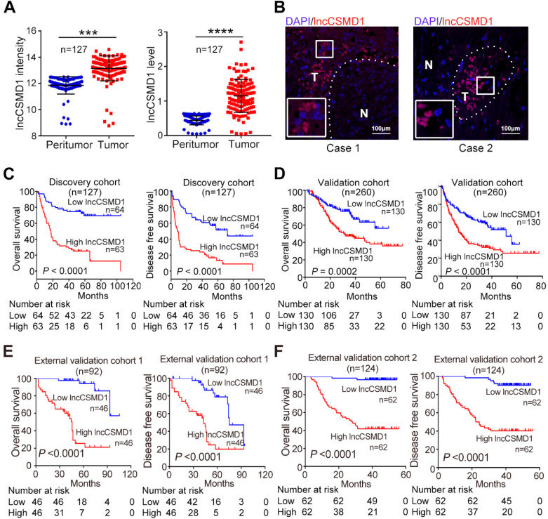
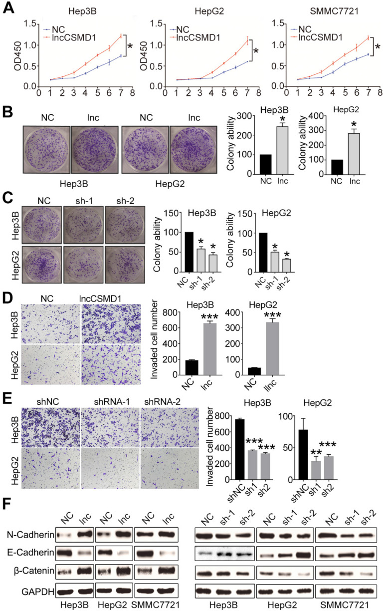
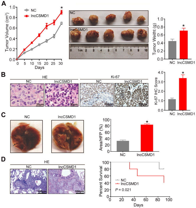
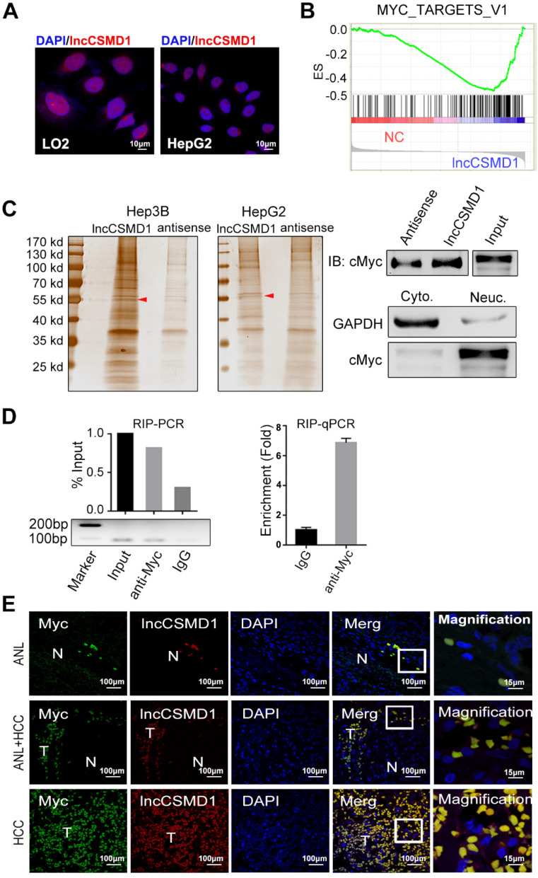
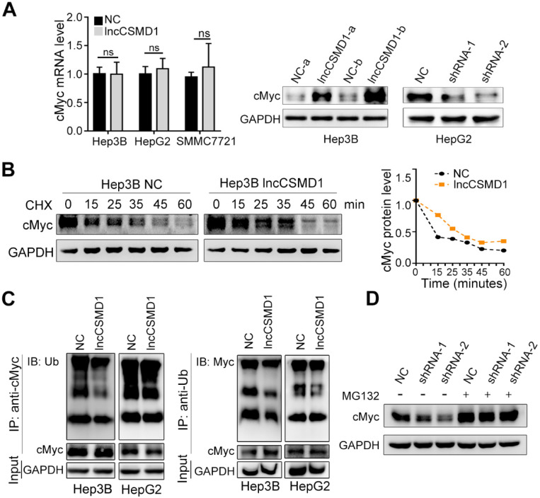
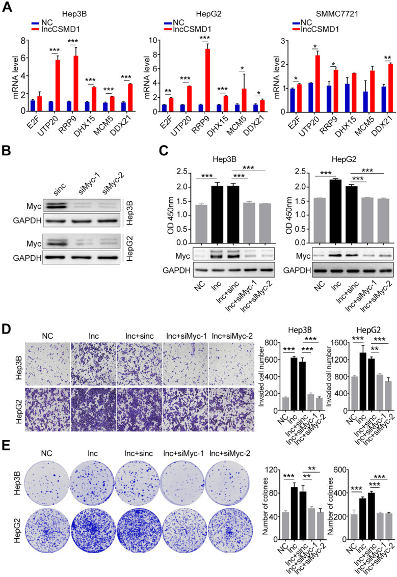
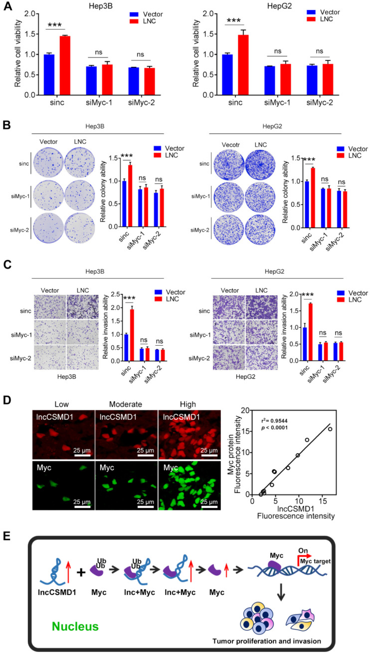
Emerging evidence suggests that long non-coding RNAs (lncRNA) play critical roles in the development and progression of diverse cancers including hepatocellular carcinoma (HCC), but the underlying molecular mechanisms of lncRNAs that are involved in hepatocarcinogenesis have not been fully explored. Methods: In this study, we profiled lncRNA expression in 127 pairs of HCC and nontumor liver tissues (a Discovery Cohort) using a custom microarray. The expression and clinical significance of lncCSMD1-1 were then validated with qRT-PCR and COX regression analysis in a Validation Cohort (n=260) and two External Validation Cohorts (n=92 and n=124, respectively). In vitro and in vivo assays were performed to explore the biological effects of lncCSMD1-1 on HCC cells. The interaction of lncCSMD1-1 with MYC was identified by RNA pull-down and RNA immunoprecipitation. The role of LncCSMD1-1 in the degradation of MYC protein was also investigated. Results: With microarray, we identified a highly upregulated lncRNA, lncCSMD1-1, which was associated with tumor progression and poor prognosis in the Discovery Cohort, and validated in another 3 HCC cohorts. Consistently, ectopic expression of lncCSMD1-1 notably promotes cell proliferation, migration, invasion, tumor growth and metastasis of HCC cells in in vitro and in vivo experiments. Gene expression profiling on HCC cells and gene sets enrichment analysis indicated that the MYC target gene set was significantly enriched in HCC cells overexpressing lncCSMD1-1, and lncCSMD1-1 was found to directly bind to MYC protein in the nucleus of HCC cells, which resulted in the elevation of MYC protein. Mechanistically, lncCSMD1-1 interacted with MYC protein to block its ubiquitin-proteasome degradation pathway, leading to activation of its downstream target genes. Conclusion: lncCSMD1-1 is upregulated in HCC and promotes progression of HCC by activating the MYC signaling pathway. These results provide the evidence that lncCSMD1-1 may serve as a novel prognostic marker and potential therapeutic target for HCC.

Keywords: Hepatocellular carcinoma; Long noncoding RNA; MYC; lncCSMD1-1; prognosis.


Conflict of interest statement

Competing Interests: The authors have declared that no competing interest exists.

Figures

Figure 1
Figure 1
lncCSMD1 expression is increased and positively correlated with poor prognosis in HCC patients. (A) Relative lncCSMD1 expression level was measured in 127 paired HCC and peritumor liver tissues (Discovery Cohort) with a custom lncRNA microarray (left) and in another 127 paired HCC and peritumor liver tissues (randomly selected from the Validation Cohort) with qRT-PCR (right). (B) lncCSMD1 was mainly expressed in HCC tissues (T), but rarely in peritumor liver tissues (N), as shown by RNA fluorescence in situ hybridization. (C) In the Discovery Cohort, 127 HCC patients were divided into high or low expression group according to the median value of lncCSMD1 expression detected by custom lncRNA microarray. Kaplan-Meier analysis revealed that HCC patients with high lncCSMD1 expression level have significantly worse overall survival (OS, left) and disease-free survival (DFS, right) than those with low lncCSMD1 level. (D-F) The HCC patients in the Validation Cohort (D), External Validation Cohort 1 (E) and 2 (F) were divided into high or low expression group according to the median value of lncCSMD1 level detected by qRT-PCR. Kaplan-Meier analysis of these Validation Cohorts verified the results obtained in the Discovery Cohort.
Figure 2
Figure 2
Ectopic expression of lncCSMD1 promotes proliferation, colony formation and invasion of HCC cells in vitro. (A) The ectopic expression of lncCSMD1 markedly promotes cell proliferation in Hep3B, HepG2 and SMMC7721 HCC cells, which were determined by CCK-8 assay. (B) lncCSMD1 overexpression significantly enhances colony formation in Hep3B and HepG2 HCC cells compared with the control cells. Data represent the mean ± SD of triplicate experiments and was analyzed by Student t test. * means p <0.05. (also applies to the following). (C) Downregulated lncCSMD1 notably inhibits colony formation in Hep3B and HepG2 HCC cells compared with the control cells. (D) In transwell assay, lncCSMD1 expression strikingly raises the cell invasion in Hep3B and HepG2 cells compared with the control cells. (E) In transwell assay, downregulated lncCSMD1 by shRNA markedly reduces the cell invasion in Hep3B and HepG2 cells compared with the control cells. (F) Overexpression of lncCSMD1 causes the expression of mesenchymal proteins (N-cadherin and β-catenin) and reduces the expression of epithelial protein (E-cadherin) in Hep3B, HepG2 and SMMC7721 cells, as shown by western blot.
Figure 3
Figure 3
LncCSMD1 promotes growth and metastasis of xenograft tumors derived from Hep3B HCC cells. (A) In the tumor growth curve, the tumors grow faster in the mice subcutaneously inoculated with Hep3B cells overexpressing lncCSMD1 than in mice with the HCC cells carrying a control vector (left); Images of xenograft tumors derived from Hep3B cells stably overexpressing lncCSMD1 or carrying a control vector (middle); the weights of xenograft tumors in the two groups were compared with histogram (right), in which data were analyzed by sample-paired t test; *P <0.05 (the followings are the same). (B) Representative images of HE staining (left) and Ki67 IHC staining (middle) displayed the pathological configuration and proliferation capacity of HCC xenograft cells; IHC scores for Ki67 were compared in the two groups with histogram (right). (C) Representative images of nude mouse lungs with metastatic foci derived from tail-vein injected Hep3B cells stably overexpressing lncCSMD1 or carrying a control vector (n=5 mice/each group); the metastatic foci ratios (foci to the observed lung area) were compared in the two groups with histogram. (D) Representative images of metastatic foci stained with HE in the lung tissue sections (left); survivals mice tail-vein injected with Hep3B cells stably expressing lncCSMD1 or a control vector were analyzed with Kaplan-Meier curve (right).
Figure 4
Figure 4
LncCSMD1 directly interacts with MYC protein. (A) LncCSMD1 is located in the nucleus of HepG2 and LO2 cells, as shown by RNA-FISH assay; (B) Gene set enrichment analysis (GSEA) for gene expression array data from Hep3B cells stably expressing lncCSMD1 and a control vector indicates a significant association between lncCSMD1 and MYC target gene signature. (C) Silver staining images of PAGE gels, in which lncCSMD1/proteins complexes from RNA pull down experiments on nucleus proteins of HCC cells stably expressing lncCSMD1 were separated; the arrows show the specific protein bands in pull down complexes by lncCSMD1 sense sequence when compared with antisense sequence (left); Immunoblot analysis verified the interaction between MYC and lncCSMD1 sense sequence (right). (D) LncCSMD1 PCR products amplified from RNA immunoprecipitation (RIP) complexes are separated on Agarose gel (lower left); the lncCSMD1 PCR products are quantified and compared with histogram ( upper left); quantitative PCR was used to compare the lncCSMD1 amount that binds to MYC protein in the RIP experiment (right). (E) MYC protein is overlapped with lncCSMD1 RNA in the nucleus of HCC and peritumor liver cells, as displayed by Immunofluorescence (for MYC protein) coupled with fluorescence in situ hybridization (for lncCSMD1). T: HCC tissue; N: liver tissue.
Figure 5
Figure 5
LncCSMD1 stabilized MYC protein by inhibiting its ubiquitination. (A) MYC mRNA expression level in Hep3B cells stably expressing lncCSMD1 is not significantly higher than that in the control cells, as exhibited by qRT-PCR (left); however, MYC protein level is much higher in Hep3B cells stable overexpression of lncCSMD1 than in control cells (middle) or obviously lower in HepG2 cells with lncCSMD1 downregulation than in control cells (right). (B) Hep3B cells were treated with cycloheximide (CHX; 20 μg/ml) for the indicated times, and the half-life of MYC protein in HCC cells with overexpressing lncCSMD1 is prolonged when compared with that in control cells, as revealed by immunobloting (left); the curves were used to compare the half-life times of MYC protein in Hep3B cells with overexpression of lncCSMD1 or vector (right). (C) HCC cells overexpressing lncCSMD1 or vector were treated with MG132 (5 μM) for 24 h, and then cell lysates were immunoprecipitated with MYC antibody and the precipitated complexes were subjected to western blot with ubiquitin antibody (left); or the cell lysates were immunoprecipitated with ubiquitin antibody and the precipitated complexes were subjected to western blot with MYC antibody (right). The two experiments are aimed to detect the ubiquitination status of MYC protein. (D) MYC protein in Hep3B cells with downregulation of lncCSMD1 by shRNA is reduced compared with that in negative control cells, while after MG132 (20 μM) treatment for 24 h, MYC protein is restored to the same level as that in control cells, as shown by western blot assay.
Figure 6
Figure 6
MYC protein plays a key role in lncCSMD1-induced malignant phenotypes of HCC cells. (A) MYC target genes in Hep3B, HepG2 and SMMC7721 cells with overexpression of lncCSMD1 are expressed higher than those in the control cells, as shown by qRT-PCR. (B) MYC protein expression is reduced by siRNA against MYC in Hep3B and HepG2 cells, as shown by Western Blot. (C) MYC protein in Hep3B and HepG2 cells with lncCSMD1 overexpression is elevated, as displayed by western blot (lower panels) and promotes cell proliferation as shown by CCK-8 assay (upper panels) compared with that in control cells; when the HCC cells were treated with siMYC again, MYC protein was reduced and leads to decrease in cell proliferation. (D, E) The same treatments as in the above (C) were conducted in the same Hep3B and HepG2 cells, and these cells were determined with transwell assay (D) and colony formation assay (E), in which overexpressed lncCSMD1 promotes the invasion and colony formation of the HCC cells and the downregulated MYC by siRNA inhibits colony formation and invasion of the HCC cells.
Figure 7
Figure 7
LncCSMD1 failed to induce malignant phenotypes of HCC cells under knock-down of MYC. (A-C) In Hep3B and HepG2 cells, MYC was knocked down by siRNA against MYC followed by transient overexpression of lncCSMD1, and then CCK8 (A), colony formation (B) and invasion assay (C) were carried out to evaluate the effect of lncCSMD1 overexpression on the proliferation, colony formation and invasion ability of these HCC cells, showing that lncCSMD1 overexpression can promote proliferation, colony formation and invasion of the control HCC cells, but has not any significant effect on the biological phenotypes of the HCC cells with downregulation of MYC compared with those of the HCC cells with only MYC knockdown. (D) The expressions of lncCSMD1 RNA and MYC protein in 11 primary HCC tissues were investigated by FISH and immunofluorescence, respectively, and the representative images are presented (left); In Pearson correlation analysis, lncCSMD1 RNA has a highly positive relationship with MYC protein in HCC tissues (11 cases) (right). (E) The work model of lncCSMD-induced oncogenic phenotypes of HCC cells via activating MYC signaling.

Similar articles

Cited by

References

    1. Bray F, Ferlay J, Soerjomataram I, Siegel RL, Torre LA, Jemal A. Global cancer statistics 2018: GLOBOCAN estimates of incidence and mortality worldwide for 36 cancers in 185 countries. CA Cancer J Clin. 2018;68:394–424. - PubMed
    1. Omata M, Cheng AL, Kokudo N, Kudo M, Lee JM, Jia J. et al. Asia-Pacific clinical practice guidelines on the management of hepatocellular carcinoma: a 2017 update. Hepatol Int. 2017;11:317–70. - PMC - PubMed
    1. Pi J, Kang Y, Smith M, Earl M, Norigian Z, McBride A. A review in the treatment of oncologic emergencies. J Oncol Pharm Pract. 2016;22:625–38. - PubMed
    1. Dang CV, Resar LM, Emison E, Kim S, Li Q, Prescott JE. et al. Function of the c-Myc oncogenic transcription factor. Exp Cell Res. 1999;253:63–77. - PubMed
    1. Niu ZS, Li BK, Wang M. Expression of p53 and C-myc genes and its clinical relevance in the hepatocellular carcinomatous and pericarcinomatous tissues. World J Gastroenterol. 2002;8:822–6. - PMC - PubMed

Publication types

MeSH terms