. 2020 Sep;39(38):6099-6112.
doi: 10.1038/s41388-020-01417-6. Epub 2020 Aug 18.

A novel mitochondrial amidoxime reducing component 2 is a favorable indicator of cancer and suppresses the progression of hepatocellular carcinoma by regulating the expression of p27

Affiliations

A novel mitochondrial amidoxime reducing component 2 is a favorable indicator of cancer and suppresses the progression of hepatocellular carcinoma by regulating the expression of p27

Dehai Wu et al. Oncogene. 2020 Sep.

Abstract

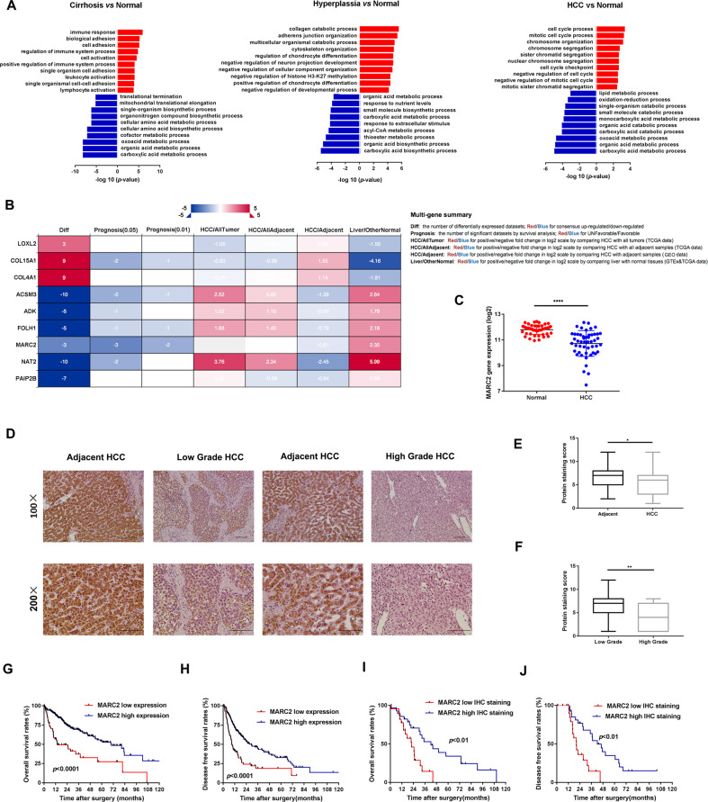
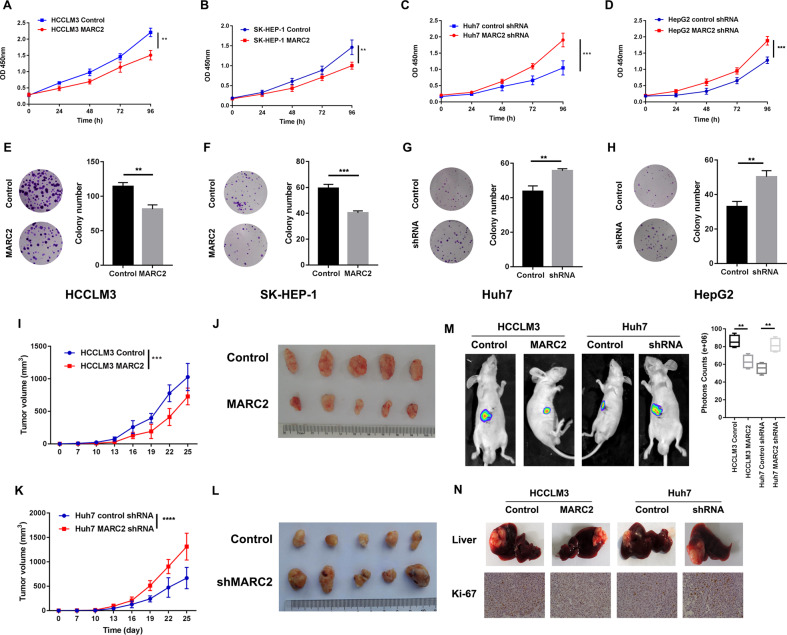
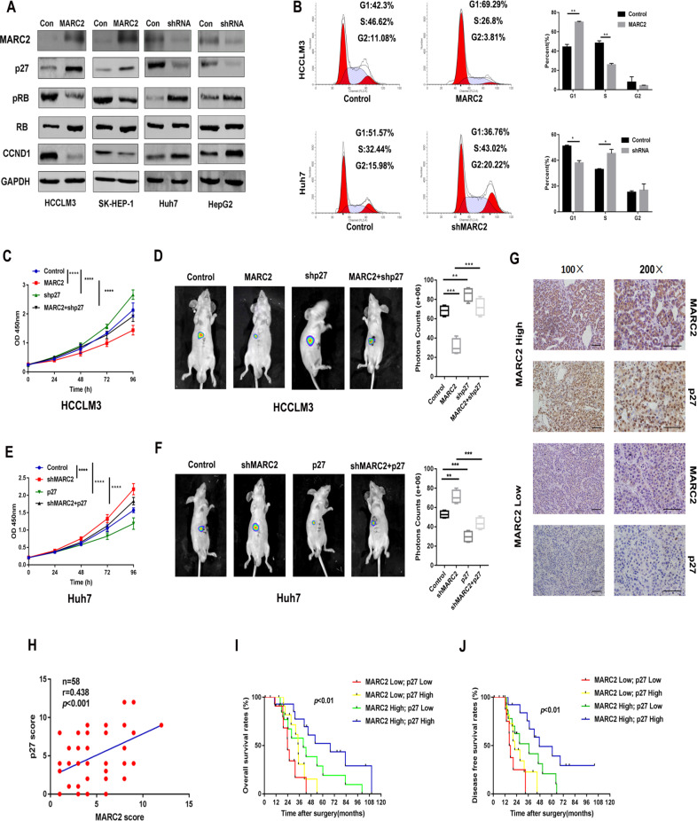
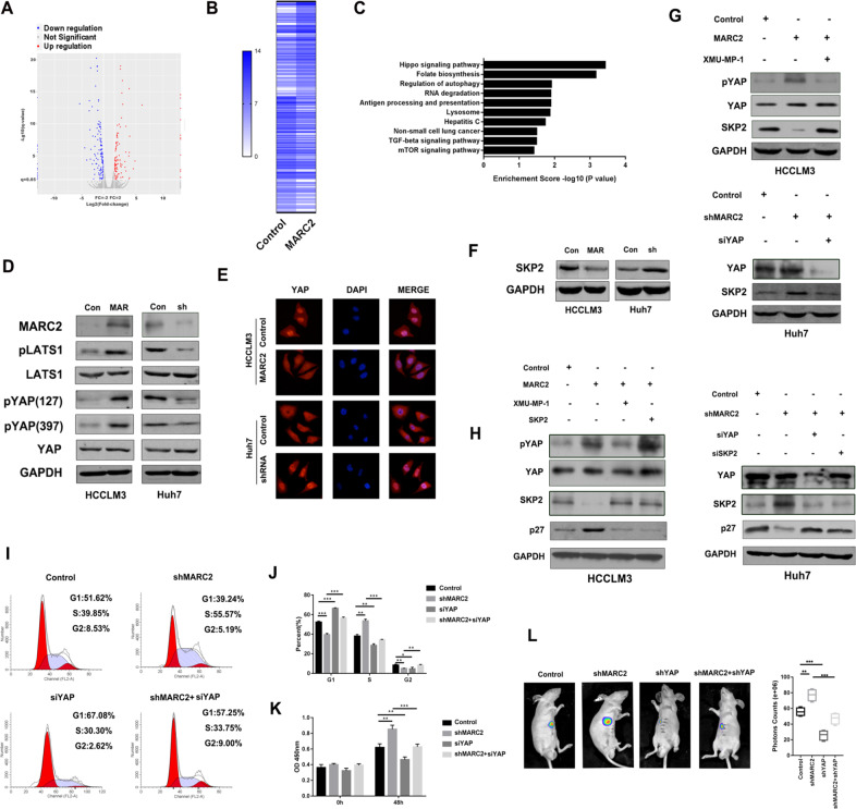
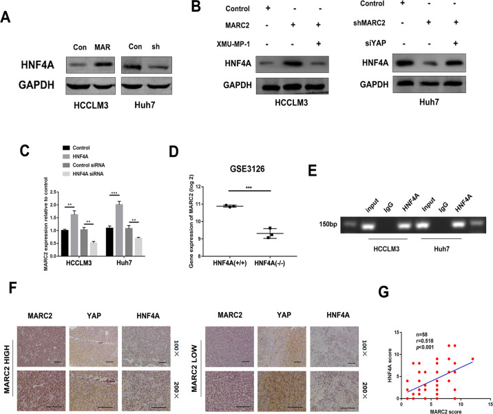
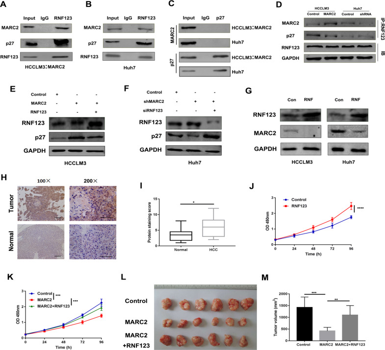
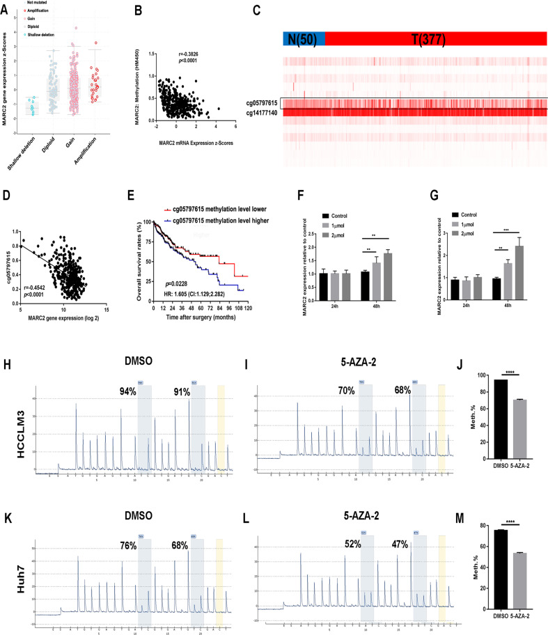
Hepatocellular carcinoma (HCC) is the fifth leading cause of cancer-related mortality in the United States. Exploring the mechanism of HCC and identifying ideal targets is critical. In the present study, we demonstrated metabolism dysfunction might be a key diver for the development of HCC. The mitochondrial amidoxime reducing component 2 (MARC2) as a newly discovered molybdenum enzyme was downregulated in human HCC tissues and HCC cells. Downregulated MARC2 was significantly associated with clinicopathological characteristics of HCC, such as tumor size, AFP levels, and tumor grade and was an independent risk factor of poor prognosis. Both in vitro and in vivo studies suggested that MARC2 suppressed the progression of HCC by regulating the protein expression level of p27. The Hippo signaling pathway and RNF123 were required for this process. Moreover, MARC2 regulated expression of HNF4A via the Hippo signaling pathway. HNF4A was recruited to the promoter of MARC2 forming a feedback loop. MARC2 levels were downregulated by methylation. We demonstrated the prognostic value of MARC2 in HCC and determined the mechanism by which MARC2 suppressed the progression of HCC in this study. These findings may lead to new therapeutic targets for HCC.


Conflict of interest statement

The authors declare that they have no conflict of interest.

Figures

Fig. 1
Fig. 1. MARC2 is downregulated in HCC and predicted poor prognosis.
a Gene expression data were extracted from GSE3500, GSE6764, and GSE14520. Different expression gene existed in all the three databases were filtered and separated into two groups (upregulated group: red and downregulated group: blue) enriching in cell process. b Filtered genes were valid in different databases in the left panel; the annotation for column in the datasheet in the right panel. The survival analysis of MARC2 was significant in three datasets (prognosis p < 0.05), and more significant in two datasets (prognosis p < 0.01). c MARC2 mRNA levels were significantly decreased in HCC tissues (n = 50) compared with the paired adjacent normal tissues (n = 50). d Representative images of MARC2 IHC staining in human HCC tissues and adjacent normal liver tissues. Bar = 100 µm (*100); Bar = 50 µm (*200). e IHC staining score was used to compare the protein expression of MARC2 between HCC tissues (n = 57) and the adjacent normal tissues (n = 82). f IHC staining score was used to compare the protein expression of MARC2 between patient with low tumor grade (n = 36) and patient with high tumor grade (n = 19). g, h The Kaplan–Meier method was used to compare the OS and DFS between the patient group with high level of MARC2 and patient group with low level of MARC2. i, j The Kaplan–Meier plots of OS and DFS between patients with high (n = 15) and low level of MARC2 (n = 15) according to IHC scores. The log-rank test was used to compare survival between different groups. *p < 0.05; **p < 0.01; ***p < 0.001, all of which were considered statistically significant.
Fig. 2
Fig. 2. MARC2 suppresses the progress of HCC both in vitro and in vivo.
ad Proliferation rate was analyzed by CCK8 assay in the indicated HCC cell lines. eh Images represented the colony formation assay in the indicated cell lines in the left panel and statistical analysis of colony numbers are in the right panel. k, i Tumor growth curves of subcutaneous tumor for the indicated cell lines. The tumor volume was measured per 3 day since the 7th day. j, l Representative images of subcutaneous xenograft derived from indicated cell lines. Five mice were used for each group. m Representative images of liver xenograft derived from indicated cell lines in left panel; the statistical analysis of bioluminescence signal in the right panel. Four mice were used for each group. n Representative images of livers (up) and IHC staining of Ki-67 (down) for the liver xenograft models derived from the indicated cell lines. The data are presented as mean ± SD. *p < 0.05; **p < 0.01; ***p < 0.001, all of which were considered statistically significant.
Fig. 3
Fig. 3. MARC2 suppresses the progression of HCC via regulating the protein expression of p27.
a Expression of MARC2 and cell cycle associated signal proteins in the indicated cell lines was measured by western blot. b Representative images of cell cycle phase in the indicated cell lines in the left panel and statistical analysis in the right panel. c, e The growth curves of the indicated cell lines representing the proliferation rates were measured by CCK8 assay. d, f Representative images of liver xenograft derived from indicated cell lines in the left panel and the statistical analysis of bioluminescence signal in the right panel. c, d HCCLM3 cell line was used for the cell and mice experiment. e, f Huh7 cell line was used for the cell and mice experiment. g IHC staining of MARC2 and p27 in human HCC tissues. Bar = 100 µm (*100); Bar = 50 µm (*200). h IHC scores were used to exam correlation of protein expression between p27 and MARC2. The Pearson correlation coefficient was used to determine the correlation. i, j The Kaplan–Meier method was used to determine the overall survival and disease free survival of patient with HCC; the log-rank test was used to compare survival between different groups. *p < 0.05; **p < 0.01; ***p < 0.001, all of which were considered statistically significant.
Fig. 4
Fig. 4. MARC2 regulates the expression of p27 via Hippo signaling pathway.
a Volcano plot for different expression genes between HCCLM3 control and HCCLM3-MARC2 cell lines. Red plots represented the upregulated genes and blue plots represented the downregulated genes. The significant threshold were |fold change| > 2 and p < 0.05. b Heatmap representing the mRNA expression of significant different expression genes between HCCLM3 control and HCCLM3-MARC2 cells. c Significant different expression genes were filtered and enriched in KEGG signaling pathway; the Top10 signaling pathways were listed and ranked by P value. d Representative the WB images for Hippo signaling pathway in indicated cell lines. Con: Control; MAR: MARC2 overexpression; sh: MARC2 shRNA. e Expression of YAP in the indicated cells was examined by immunofluorescence. Single and merged images were taken in the indicated cells to show immunofluorescence staining of YAP (red), accompanied by the cell nucleus (blue) stained by DAPI. f Expression of SKP2 was detected by western blot in indicated cell lines. g XMU-MP-1 was used to active the YAP. 3 μM XMU-MP-1 was added to MARC2-HCCLM3 cell lines, and the expression of pYAP, YAP, and SKP2 was detected by western blot in the upper panel. SiYAP was used in shMARC2-Huh7 cell lines to suppress the Hippo signaling pathway, and the expression of YAP and SKP2 was detected by western blot in the lower panel. h To further confirmed that MARC2 regulated p27 via YAP/SKP2 axis, 3 μM XMU-MP-1 and SKP2 overexpression plasmid were added to MARC2-HCCLM3 cell lines respectively, and the expression of pYAP, YAP, SKP2, and p27 was detected by western blot in the left panel. SiYAP and siSKP2 were added to shMARC2-Huh7 cell lines respectively, and the expression of YAP, SKP2, and p27 was detected by western blot in the right panel. i Representative images for cell cycle assays in indicated cell lines. j Statistical analysis of cell cycle. k Proliferation rates were measured by CCK8 assay in the indicated cell lines at 0 h and 48 h. l Representative images of liver xenograft derived from indicated cell lines in the left panel; the statistical analysis of bioluminescence signal in the right panel. il Huh7 cell line was used for the experiment. *p < 0.05; **p < 0.01; ***p < 0.001, all of which were considered statistically significant.
Fig. 5
Fig. 5. MARC2 regulates HNF4A by formation of a feedback loop via the Hippo signaling pathway.
a, b Protein expression of HNF4A was detected by western blot in indicated cell lines. XMU-MP-1 was used to active the YAP, while the siYAP was used to suppress the Hippo signaling pathway. To further confirmed regulated HNF4A via Hippo signaling pathway, XMU-MP-1 was added to HCCLM3-MARC2 cell line and siYAP was added to Huh7-shMARC2 cell line. c Gene expression of MARC2 was measured by real-time PCR in indicated cell lines. d Gene expression of MARC2 in HNF4A KO mice. Data were obtained from GSE3126. HNF4A (+/+) was used as control group and HNF4A (−/−) represented the HNF4A knockout group. e CHIP-PCR assay showed HNF4A was recruited to promoter region of MARC2. f Representative the images of IHC staining for MARC2, YAP, and HNF4A in human HCC tissues. Bar = 100 µm (*100); Bar = 50 µm (*200). g IHC scores were used to exam correlation of protein expression of HNF4A and MARC2. The Pearson correlation coefficient was used to determine the relationship of protein expression between HNF4A and MARC2. *p < 0.05; **p < 0.01; ***p < 0.001.
Fig. 6
Fig. 6. MARC2 combines with RNF123 to prevent the degradation of p27.
a, b The indicated cell lysates were prepared and immunoprecipitated with anti-RNF123 antibody. The protein expressions of MARC2, p27 and RNF123 were detected by western blot. HCCLM3 with overexpression of MARC2 represented the exogenous MARC2 interaction with RNF123, and Huh7 cell line represented the endogenous MARC2 interaction with RNF123. c The indicated cell lysates were prepared and immunoprecipitated with anti-p27 antibody. Immunoprecipitates and cell lysates were analyzed by western blot. d The indicated cell lysates were prepared and immunoprecipitated with anti-RNF123 antibody. The protein expressions of MARC2, p27, and RNF123 were detected by western blot. e, f The protein expression of RNF123 and p27 was detected by western blot in the indicated cell lines. g The protein expression of RNF123 and MARC2 was detected by western blot in the indicated cell lines. Con: Control; RNF: RNF123 overexpression. h Representative images of IHC staining for RNF123 in human HCC and adjacent normal tissues. Bar = 100 µm (*100); Bar = 50 µm (*200). i RNF123 IHC scores were used to compare the expression of RNF123 between normal tissues and HCC tissues. j, k Proliferation rate was analyzed by CCK8 assay in the indicated cell lines. l The images of subcutaneous xenograft derived from indicated cell lines. m Statistical analysis of tumor volume. jm The experiment was derived from HCCLM3 cell line. *p < 0.05; **p < 0.01; ***p < 0.001.
Fig. 7
Fig. 7. MARC2 is downregulated by methylation.
a Gene expression of MARC2 in different copy number groups. Shallow deletion: 0.5; Diploid: 1; Gain: 1.5; Amplification: 2. b Correlation between MARC2 gene expression and methylation score of MARC2. The Pearson correlation coefficient was used to determine the relationship. c Heatmap for the CpG sites of MARC2. N represented the normal tissue (n = 50), and T represented the tumor tissue (n = 377). d Correlation between MARC2 gene expression and methylation score of cg05797615. The Pearson correlation coefficient was used to determine the relationship. e The Kaplan–Meier method was used to compare the OS between the patient group with high methylation score of cg05797615 and patient group with low methylation score of cg05797615. f, g 0, 1, 2 µmol 5-Aza-2′-deoxycytidine was added to HCCLM3 and Huh7 cell lines respectively. qPCR was used to analyze the gene expression of MARC2 in HCCLM3 and Huh7 cell lines treated with 5-Aza-2′-deoxycytidine at 24 h and 48 h. hm Pyrosequencing was used to analyze the methylation level of cg05797615 in HCCLM3 and Huh7 cell lines treated with 5-Aza-2′-deoxycytidine in the left panel and statistical analysis in the right panel. *p < 0.05; **p < 0.01; ***p < 0.001.

Similar articles

Cited by

References

    1. Siegel RL, Miller KD, Jemal A. Cancer statistics, 2020. CA Cancer J Clin. 2020;70:7–30. - PubMed
    1. Massarweh NN, El-Serag HB. Epidemiology of hepatocellular carcinoma and intrahepatic cholangiocarcinoma. Cancer Control. 2017;24:1073274817729245. doi: 10.1177/1073274817729245. - DOI - PMC - PubMed
    1. Guichard C, Amaddeo G, Imbeaud S, Ladeiro Y, Pelletier L, Maad IB, et al. Integrated analysis of somatic mutations and focal copy-number changes identifies key genes and pathways in hepatocellular carcinoma. Nat Genet. 2012;44:694–8. doi: 10.1038/ng.2256. - DOI - PMC - PubMed
    1. Wang X, Chen D, Chen B. The long-to-short-axis ratio and multifocality are associated with TP53 mutation status in surgically resected hepatocellular carcinomas. Acad Radiol. 2018; e-pub ahead of print 27 June 2018; 10.1016/j.acra.2018.04.021. - PubMed
    1. Wahl B, Reichmann D, Niks D, Krompholz N, Havemeyer A, Clement B, et al. Biochemical and spectroscopic characterization of the human mitochondrial amidoxime reducing components hmARC-1 and hmARC-2 suggests the existence of a new molybdenum enzyme family in eukaryotes. J Biol Chem. 2010;285:37847–59. doi: 10.1074/jbc.M110.169532. - DOI - PMC - PubMed

MeSH terms